

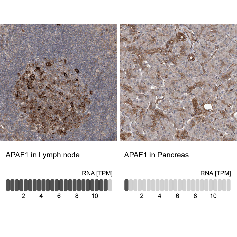
Immunohistochemistry analysis in human lymph node and pancreas tissues using HPA031373 antibody. Corresponding APAF1 RNA-seq data are presented for the same tissues.
Immunohistochemistry analysis in human lymph node and pancreas tissues using HPA031373 antibody. Corresponding APAF1 RNA-seq data are presented for the same tissues.
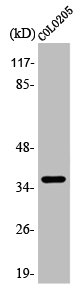
Anti-APAF1 Antibody
HPA031373
ApplicationsImmunoHistoChemistry
Product group Antibodies
ReactivityHuman
TargetAPAF1
Overview
- SupplierAtlas Antibodies
- Product NameAnti-APAF1 Antibody
- Delivery Days Customer4
- ApplicationsImmunoHistoChemistry
- CertificationResearch Use Only
- ClonalityPolyclonal
- ConjugateUnconjugated
- Gene ID317
- Target nameAPAF1
- Target descriptionapoptotic peptidase activating factor 1
- Target synonymsAPAF-1, CED4, apoptotic protease-activating factor 1
- HostRabbit
- IsotypeIgG
- Protein IDO14727
- Protein NameApoptotic protease-activating factor 1
- Scientific DescriptionRecombinant Protein Epitope Signature Tag (PrEST) antigen sequence
- ReactivityHuman
- Storage Instruction-20°C,2°C to 8°C
- UNSPSC41116161
Datasheet
MSDS
Related products
Product group Antibodies
Anti-APAF1 AntibodyA97715
ApplicationsWestern Blot, ELISA
ReactivityHuman, Mouse, Rat
- SizePrice
Product group Antibodies
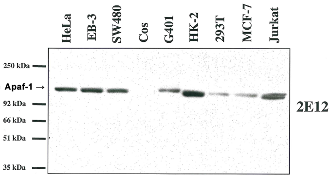
anti-Apaf-1 (human), mAb (2E12)AG-20T-0132
ApplicationsImmunoPrecipitation, Western Blot, ELISA, ImmunoCytoChemistry, ImmunoHistoChemistry
ReactivityHuman
TargetAPAF1
- SizePrice

Product group Antibodies
Anti-APAF1 Antibody Picoband(r)A00889-2-CARRIER-FREE
ApplicationsFlow Cytometry, Western Blot, ELISA
ReactivityHuman
TargetAPAF1
- SizePrice
Product group Antibodies

APAF1 AntibodyABX324778
ApplicationsImmunoFluorescence, Western Blot, ELISA, ImmunoCytoChemistry, ImmunoHistoChemistry
- SizePrice
Product group Antibodies
References
APAF1(CT) Polyclonal AntibodyBS-0058R
ApplicationsImmunoFluorescence, Western Blot, ELISA, ImmunoCytoChemistry, ImmunoHistoChemistry, ImmunoHistoChemistry Frozen, ImmunoHistoChemistry Paraffin
ReactivityCanine, Equine, Human, Mouse, Rat
TargetAPAF1
- SizePrice
Product group Antibodies
APAF1 AntibodyCSB-PA000895
ApplicationsImmunoFluorescence, Western Blot, ELISA, ImmunoHistoChemistry
ReactivityHuman
- SizePrice
Product group Antibodies
Apaf1 Polyclonal AntibodyCAC08235
ApplicationsImmunoFluorescence, ELISA, ImmunoHistoChemistry
TargetAPAF1
- SizePrice
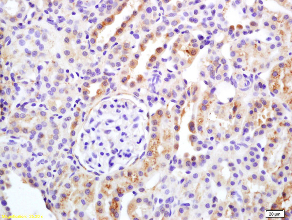
![APAF1 antibody [C3], C-term detects APAF1 protein at cytoplasm in rat liver by immunohistochemical analysis. Sample: Paraffin-embedded rat liver. APAF1 antibody [C3], C-term (GTX101067) diluted at 1:400.
Antigen Retrieval: Citrate buffer, pH 6.0, 15 min](https://www.genetex.com/upload/website/prouct_img/normal/GTX101067/GTX101067_40359_20160713_IHC-P_R_w_23060100_579.webp)
Product group Antibodies
APAF1 antibody [C3], C-termGTX101067
ApplicationsImmunoHistoChemistry, ImmunoHistoChemistry Paraffin
ReactivityHuman, Mouse, Rat
TargetAPAF1
- SizePrice