

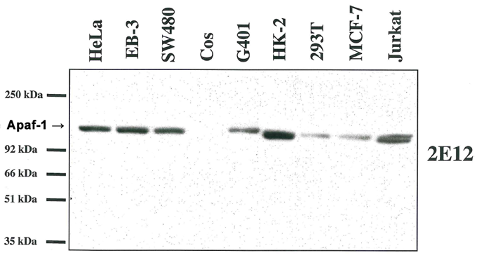
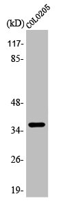
Western blot analysis using anti-Apaf-1 (human), MAb (2E12) (AG-20T-0132) on several human cell lines and one monkey cell line (COS).
Western blot analysis using anti-Apaf-1 (human), MAb (2E12) (AG-20T-0132) on several human cell lines and one monkey cell line (COS).
anti-Apaf-1 (human), mAb (2E12)
AG-20T-0132
ApplicationsImmunoPrecipitation, Western Blot, ELISA, ImmunoCytoChemistry, ImmunoHistoChemistry
Product group Antibodies
ReactivityHuman
TargetAPAF1
Overview
- SupplierAdipoGen Life Sciences
- Product Nameanti-Apaf-1 (human), mAb (2E12)
- Delivery Days Customer10
- ApplicationsImmunoPrecipitation, Western Blot, ELISA, ImmunoCytoChemistry, ImmunoHistoChemistry
- CertificationResearch Use Only
- ClonalityMonoclonal
- Clone ID2E12
- Concentration1 mg/ml
- Estimated Purity>95%
- Gene ID317
- Target nameAPAF1
- Target descriptionapoptotic peptidase activating factor 1
- Target synonymsAPAF-1, CED4, apoptotic protease-activating factor 1
- HostRat
- IsotypeIgG2a
- Protein IDO14727
- Protein NameApoptotic protease-activating factor 1
- Scientific DescriptionApaf-1 is a cytoplasmic protein that forms one of the central hubs in the apoptosis regulatory network. It contains (from the N terminal) a caspase recruitment domain (CARD), an ATPase domain (NB-ARC), few short helical domains and then several copies of the WD40 repeat domain. Upon binding cytochrome c and dATP, this protein forms an oligomeric apoptosome, mediates the cytochrome c-dependent autocatalytic activation of pro-caspase-9 (Apaf-3), leading to the activation of caspase-3 and apoptosis. - Monoclonal Antibody. Recognizes the CARD domain of human Apaf-1. Does not cross-react with mouse or rat Apaf-1. Isotype: Rat IgG2akappa. Clone: 2.00E+12. Applications: ELISA, ICC, IHC, IP, WB. Liquid. In PBS containing 0.02% sodium azide. Apaf-1 is a cytoplasmic protein that forms one of the central hubs in the apoptosis regulatory network. It contains (from the N terminal) a caspase recruitment domain (CARD), an ATPase domain (NB-ARC), few short helical domains and then several copies of the WD40 repeat domain. Upon binding cytochrome c and dATP, this protein forms an oligomeric apoptosome, mediates the cytochrome c-dependent autocatalytic activation of pro-caspase-9 (Apaf-3), leading to the activation of caspase-3 and apoptosis.
- ReactivityHuman
- Storage Instruction-20°C,2°C to 8°C
- UNSPSC41116161
MSDS
Related products
Product group Antibodies
Anti-APAF1 AntibodyA97715
ApplicationsWestern Blot, ELISA
ReactivityHuman, Mouse, Rat
- SizePrice
Product group Antibodies
anti-Apaf-1 (mouse/rat), mAb (13F11)AG-20T-0133
ApplicationsImmunoPrecipitation, Western Blot, ELISA, ImmunoCytoChemistry
ReactivityMouse, Rat
TargetApaf1
- SizePrice
Product group Antibodies
anti-Apaf-1, mAb (18H2)AG-20T-0134
ApplicationsImmunoPrecipitation, Western Blot, ELISA, ImmunoCytoChemistry
ReactivityHuman, Mouse
TargetApaf1
- SizePrice

Product group Antibodies
Anti-APAF1 Antibody Picoband(r)A00889-2-CARRIER-FREE
ApplicationsFlow Cytometry, Western Blot, ELISA
ReactivityHuman
TargetAPAF1
- SizePrice
Product group Antibodies

APAF1 AntibodyABX324778
ApplicationsImmunoFluorescence, Western Blot, ELISA, ImmunoCytoChemistry, ImmunoHistoChemistry
- SizePrice
Product group Antibodies
References
APAF1(CT) Polyclonal AntibodyBS-0058R
ApplicationsImmunoFluorescence, Western Blot, ELISA, ImmunoCytoChemistry, ImmunoHistoChemistry, ImmunoHistoChemistry Frozen, ImmunoHistoChemistry Paraffin
ReactivityCanine, Equine, Human, Mouse, Rat
TargetAPAF1
- SizePrice
Product group Antibodies
APAF1 AntibodyCSB-PA000895
ApplicationsImmunoFluorescence, Western Blot, ELISA, ImmunoHistoChemistry
ReactivityHuman
- SizePrice
Product group Antibodies
Apaf1 Polyclonal AntibodyCAC08235
ApplicationsImmunoFluorescence, ELISA, ImmunoHistoChemistry
TargetAPAF1
- SizePrice
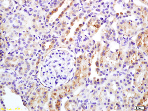
![APAF1 antibody [C3], C-term detects APAF1 protein at cytoplasm in rat liver by immunohistochemical analysis. Sample: Paraffin-embedded rat liver. APAF1 antibody [C3], C-term (GTX101067) diluted at 1:400.
Antigen Retrieval: Citrate buffer, pH 6.0, 15 min](https://www.genetex.com/upload/website/prouct_img/normal/GTX101067/GTX101067_40359_20160713_IHC-P_R_w_23060100_579.webp)
Product group Antibodies
APAF1 antibody [C3], C-termGTX101067
ApplicationsImmunoHistoChemistry, ImmunoHistoChemistry Paraffin
ReactivityHuman, Mouse, Rat
TargetAPAF1
- SizePrice
Product group Antibodies
Anti-APAF1 AntibodyHPA031373
ApplicationsImmunoHistoChemistry
ReactivityHuman
TargetAPAF1
- SizePrice