

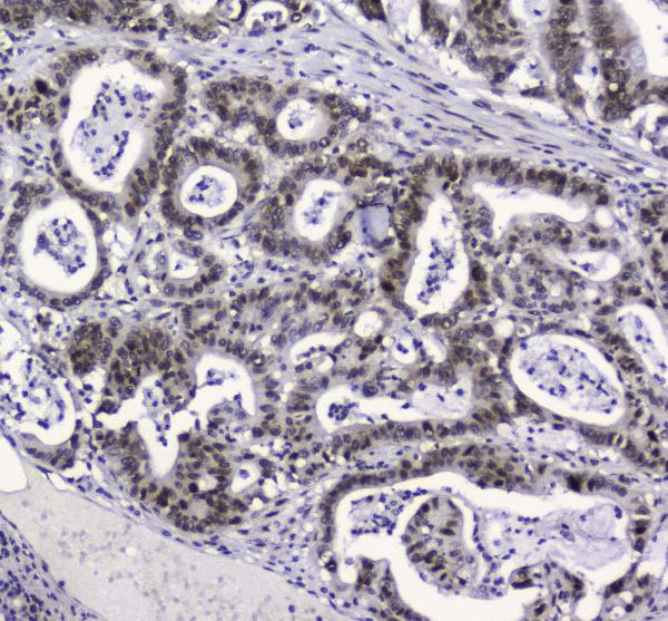
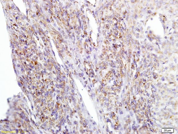
Figure 2. IHC analysis of APE1 using anti-APE1 antibody (M00627). APE1 was detected in paraffin-embedded section of human intestinal cancer tissue. Heat mediated antigen retrieval was performed in citrate buffer (pH6, epitope retrieval solution) for 20 mins. The tissue section was blocked with 10% goat serum. The tissue section was then incubated with 2microg/ml mouse anti-APE1 Antibody (M00627) overnight at 4°C. Biotinylated goat anti-mouse IgG was used as secondary antibody and incubated for 30 minutes at 37°C. The tissue section was developed using Strepavidin-Biotin-Complex (SABC)(Catalog # SA1021) with DAB as the chromogen.
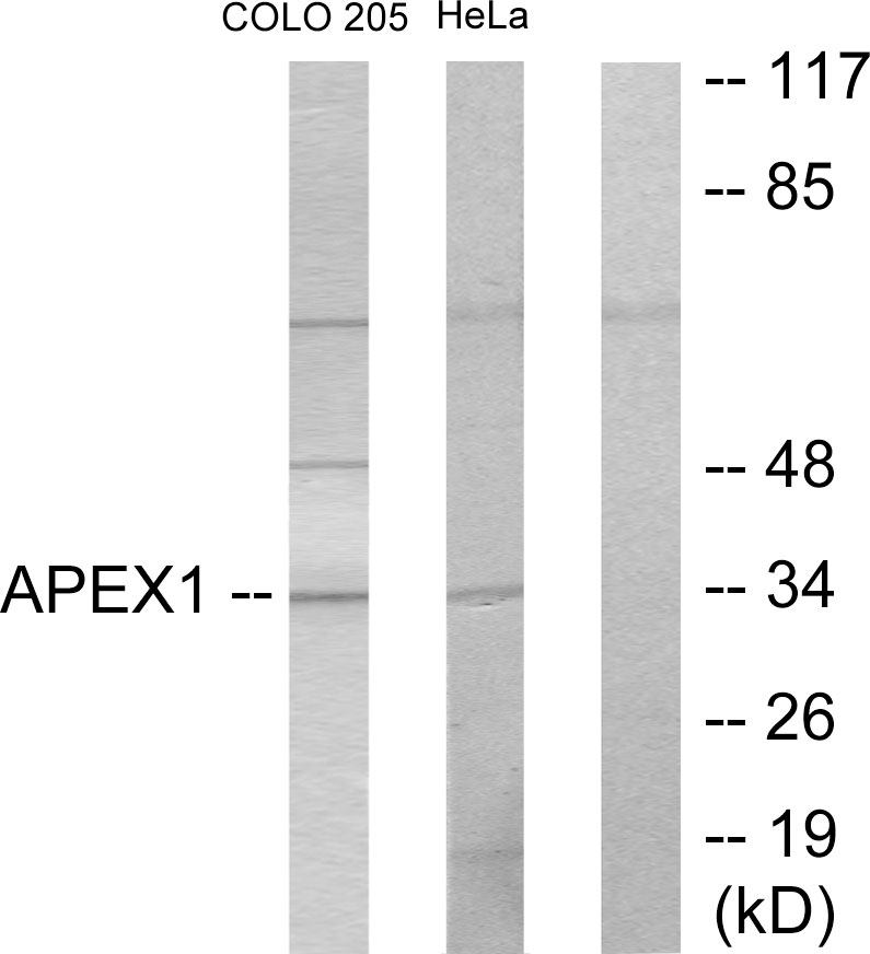
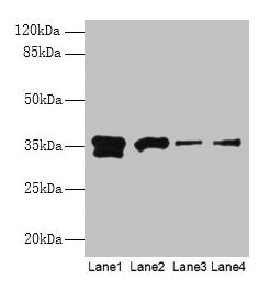
. Electrophoresis was performed on a 5-20% SDS-PAGE gel at 70V (Stacking gel) / 90V (Resolving gel) for 2-3 hours. The sample well of each lane was loaded with 50ug of sample under reducing conditions. Lane 1: human Hela whole cell lysates, Lane 2: human MCF-7 whole cell lysates, Lane 3: human COLO-320 whole cell lysates, Lane 4: human HepG2 whole cell lysates, Lane 5: Rabbit IgG, Lane 6: Marker 1113, Lane 7: human A549 whole cell lysates , Lane 8: human PANC-1 whole cell lysates, Lane 9: human 22RV1 whole cell lysates, Lane 10: human MDA-MB-453 whole cell lysates. After Electrophoresis, proteins were transferred to a Nitrocellulose membrane at 150mA for 50-90 minutes. Blocked the membrane with 5% Non-fat Milk/ TBS for 1.5 hour at RT. The membrane was incubated with mouse anti-APE1 antigen affinity purified monoclonal antibody (Catalog # M00627) at 0.5 microg/mL overnight at 4°C, then washed with TBS-0.1%Tween 3 times with 5 minutes each and probed with a goat anti-mouse IgG-HRP secondary antibody at a dilution of 1:10000 for 1.5 hour at RT. The signal is developed using an Enhanced Chemiluminescent detection (ECL) kit (Catalog # EK1001) with Tanon 5200 system.")
. Overlay histogram showing SiHa cells stained with M00627 (Blue line). To facilitate intracellular staining, cells were fixed with 4% paraformaldehyde and permeabilized with permeabilization buffer. The cells were blocked with 10% normal goat serum. And then incubated with mouse anti-APE1 Antibody (M00627, 1 microg/1x106 cells) for 30 min at 20°C. DyLight®488 conjugated goat anti-mouse IgG (BA1126, 5-10 microg/1x106 cells) was used as secondary antibody for 30 minutes at 20°C. Isotype control antibody (Green line) was mouse IgG (1 microg/1x106) used under the same conditions. Unlabelled sample without incubation with primary antibody and secondary antibody (Red line) was used as a blank control.")
Figure 2. IHC analysis of APE1 using anti-APE1 antibody (M00627). APE1 was detected in paraffin-embedded section of human intestinal cancer tissue. Heat mediated antigen retrieval was performed in citrate buffer (pH6, epitope retrieval solution) for 20 mins. The tissue section was blocked with 10% goat serum. The tissue section was then incubated with 2microg/ml mouse anti-APE1 Antibody (M00627) overnight at 4°C. Biotinylated goat anti-mouse IgG was used as secondary antibody and incubated for 30 minutes at 37°C. The tissue section was developed using Strepavidin-Biotin-Complex (SABC)(Catalog # SA1021) with DAB as the chromogen.
Anti-APE1 APEX1 Antibody Picoband(r) (monoclonal, 5C11)
M00627
ApplicationsFlow Cytometry, Western Blot, ImmunoCytoChemistry, ImmunoHistoChemistry
Product group Antibodies
ReactivityHuman
TargetAPEX1
Overview
- SupplierBoster Bio
- Product NameAnti-APE1 APEX1 Antibody Picoband(r) (monoclonal, 5C11)
- Delivery Days Customer9
- ApplicationsFlow Cytometry, Western Blot, ImmunoCytoChemistry, ImmunoHistoChemistry
- CertificationResearch Use Only
- ClonalityMonoclonal
- Clone ID5C11
- Concentration500 ug/ml
- Gene ID328
- Target nameAPEX1
- Target descriptionapurinic/apyrimidinic endodeoxyribonuclease 1
- Target synonymsAPE, APE1, APEN, APEX, APX, HAP1, REF1, DNA repair nuclease/redox regulator APEX1, AP endonuclease class I, AP lyase, APEX nuclease (multifunctional DNA repair enzyme) 1, DNA-(apurinic or apyrimidinic site) endonuclease, DNA-(apurinic or apyrimidinic site) lyase, apurinic-apyrimidinic endonuclease 1, apurinic/apyrimidinic (abasic) endonuclease, deoxyribonuclease (apurinic or apyrimidinic), protein REF-1, redox factor-1
- HostMouse
- IsotypeIgG2b
- Protein IDP27695
- Protein NameDNA repair nuclease/redox regulator APEX1
- Scientific DescriptionBoster Bio Anti-APE1 APEX1 Antibody Picoband® (monoclonal, 5C11) catalog # M00627. Tested in Flow Cytometry, IHC, ICC, WB applications. This antibody reacts with Human. The brand Picoband indicates this is a premium antibody that guarantees superior quality, high affinity, and strong signals with minimal background in Western blot applications. Only our best-performing antibodies are designated as Picoband, ensuring unmatched performance.
- ReactivityHuman
- Storage Instruction-20°C,2°C to 8°C
- UNSPSC12352203
Datasheet
MSDS
Related products
Product group Antibodies
Anti-APEX1 AntibodyA97712
ApplicationsWestern Blot, ELISA
ReactivityHuman, Mouse, Rat
- SizePrice
Product group Antibodies
Anti-APEX1 Antibody144-01117
ApplicationsImmunoFluorescence, Western Blot, ImmunoHistoChemistry
ReactivityHuman
TargetAPEX1
- SizePrice
Product group Antibodies
Girdin Polyclonal AntibodyBS-5150R
ApplicationsImmunoFluorescence, Western Blot, ELISA, ImmunoCytoChemistry, ImmunoHistoChemistry, ImmunoHistoChemistry Frozen, ImmunoHistoChemistry Paraffin
ReactivityCanine, Equine, Human, Mouse, Rabbit, Rat
TargetAPEX1
- SizePrice
Product group Antibodies
APEX1 AntibodyCSB-PA001900HA01HU
ApplicationsWestern Blot, ChIP Chromatin ImmunoPrecipitation, ELISA, ImmunoHistoChemistry
ReactivityHuman, Mouse
TargetAPEX1
- SizePrice
Product group Antibodies
References

Goat anti-APE1 / APEX1EB05345
ApplicationsWestern Blot, ELISA, ImmunoHistoChemistry
ReactivityBovine, Canine, Human, Porcine
TargetAPEX1
- SizePrice
Product group Antibodies
Apex1 Polyclonal AntibodyCAC07408
ApplicationsWestern Blot, ChIP Chromatin ImmunoPrecipitation, ELISA, ImmunoHistoChemistry
ReactivityMouse
TargetAPEX1
- SizePrice
Product group Antibodies
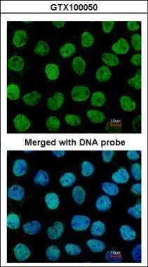
APE1 antibody [N1], N-termGTX100050
ApplicationsImmunoFluorescence, Western Blot, ImmunoCytoChemistry, ImmunoHistoChemistry, ImmunoHistoChemistry Paraffin
ReactivityHuman
TargetAPEX1
- SizePrice

Product group Antibodies
Anti-APEX1 AntibodyHPA000956
ApplicationsImmunoCytoChemistry
ReactivityHuman
TargetAPEX1
- SizePrice
Product group Antibodies

APEX1 / APE1 AntibodyLS-C400548
ApplicationsWestern Blot, ELISA, ImmunoHistoChemistry
ReactivityHuman, Mouse, Rat
TargetAPEX1
- SizePrice