

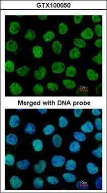
Immunofluorescence analysis of paraformaldehyde-fixed A431, using APE1(GTX100050) antibody at 1:200 dilution.
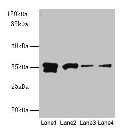
![Non-transfected (–) and transfected (+) HeLa whole cell extracts (30 μg) were separated by 10% SDS-PAGE, and the membrane was blotted with APE1 antibody [N1], N-term (GTX100050) diluted at 1:2000.](https://www.genetex.com/upload/website/prouct_img/normal/GTX100050/GTX100050_39855_20160908_WB_shRNA_watermark_w_23053123_562.webp "Non-transfected (–) and transfected (+) HeLa whole cell extracts (30 μg) were separated by 10% SDS-PAGE, and the membrane was blotted with APE1 antibody [N1], N-term (GTX100050) diluted at 1:2000.")
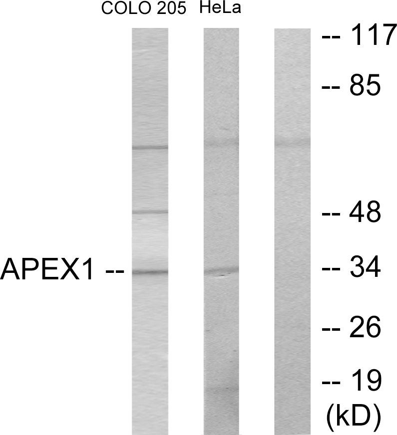
 A: A549 10% SDS PAGE GTX100050 diluted at 1:3000")
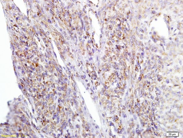
![APE1 antibody [N1], N-term detects APEX1 protein at nucleus on Saos2 xenograft by immunohistochemical analysis. Sample: Paraffin-embedded Saos2 xenograft. APE1 antibody [N1], N-term (GTX100050) dilution: 1:500.
Antigen Retrieval: Trilogy? (EDTA based, pH 8.0) buffer, 15min](https://www.genetex.com/upload/website/prouct_img/normal/GTX100050/GTX100050_39855_IHC_w_23053123_449.webp "APE1 antibody [N1], N-term detects APEX1 protein at nucleus on Saos2 xenograft by immunohistochemical analysis. Sample: Paraffin-embedded Saos2 xenograft. APE1 antibody [N1], N-term (GTX100050) dilution: 1:500.
Antigen Retrieval: Trilogy? (EDTA based, pH 8.0) buffer, 15min")
Immunofluorescence analysis of paraformaldehyde-fixed A431, using APE1(GTX100050) antibody at 1:200 dilution.
APE1 antibody [N1], N-term
GTX100050
ApplicationsImmunoFluorescence, Western Blot, ImmunoCytoChemistry, ImmunoHistoChemistry, ImmunoHistoChemistry Paraffin
Product group Antibodies
ReactivityHuman
TargetAPEX1
Overview
- SupplierGeneTex
- Product NameAPE1 antibody [N1], N-term
- Delivery Days Customer9
- Application Supplier NoteWB: 1:500-1:3000. ICC/IF: 1:100-1:1000. IHC-P: 1:100-1:1000. *Optimal dilutions/concentrations should be determined by the researcher.Not tested in other applications.
- ApplicationsImmunoFluorescence, Western Blot, ImmunoCytoChemistry, ImmunoHistoChemistry, ImmunoHistoChemistry Paraffin
- CertificationResearch Use Only
- ClonalityPolyclonal
- Concentration5.04 mg/ml
- ConjugateUnconjugated
- Gene ID328
- Target nameAPEX1
- Target descriptionapurinic/apyrimidinic endodeoxyribonuclease 1
- Target synonymsAPE, APE1, APEN, APEX, APX, HAP1, REF1, DNA repair nuclease/redox regulator APEX1, AP endonuclease class I, AP lyase, APEX nuclease (multifunctional DNA repair enzyme) 1, DNA-(apurinic or apyrimidinic site) endonuclease, DNA-(apurinic or apyrimidinic site) lyase, apurinic-apyrimidinic endonuclease 1, apurinic/apyrimidinic (abasic) endonuclease, deoxyribonuclease (apurinic or apyrimidinic), protein REF-1, redox factor-1
- HostRabbit
- IsotypeIgG
- Protein IDP27695
- Protein NameDNA repair nuclease/redox regulator APEX1
- Scientific DescriptionApurinic/apyrimidinic (AP) sites occur frequently in DNA molecules by spontaneous hydrolysis, by DNA damaging agents or by DNA glycosylases that remove specific abnormal bases. AP sites are pre-mutagenic lesions that can prevent normal DNA replication so the cell contains systems to identify and repair such sites. Class II AP endonucleases cleave the phosphodiester backbone 5 to the AP site. This gene encodes the major AP endonuclease in human cells. Splice variants have been found for this gene; all encode the same protein. [provided by RefSeq]
- ReactivityHuman
- Storage Instruction-20°C or -80°C,2°C to 8°C
- UNSPSC41116161
Datasheet
Related products
Product group Antibodies
APEX1 AntibodyCSB-PA001900HA01HU
ApplicationsWestern Blot, ChIP Chromatin ImmunoPrecipitation, ELISA, ImmunoHistoChemistry
ReactivityHuman, Mouse
TargetAPEX1
- SizePrice
Product group Antibodies
Anti-APEX1 AntibodyA97712
ApplicationsWestern Blot, ELISA
ReactivityHuman, Mouse, Rat
- SizePrice
Product group Antibodies
References

Goat anti-APE1 / APEX1EB05345
ApplicationsWestern Blot, ELISA, ImmunoHistoChemistry
ReactivityBovine, Canine, Human, Porcine
TargetAPEX1
- SizePrice

Product group Antibodies
Anti-APEX1 AntibodyHPA000956
ApplicationsImmunoCytoChemistry
ReactivityHuman
TargetAPEX1
- SizePrice
Product group Antibodies

APEX1 / APE1 AntibodyLS-C400548
ApplicationsWestern Blot, ELISA, ImmunoHistoChemistry
ReactivityHuman, Mouse, Rat
TargetAPEX1
- SizePrice
Product group Antibodies
Apex1 Polyclonal AntibodyCAC07408
ApplicationsWestern Blot, ChIP Chromatin ImmunoPrecipitation, ELISA, ImmunoHistoChemistry
ReactivityMouse
TargetAPEX1
- SizePrice

Product group Antibodies
Anti-APE1/APEX1 Antibody Picoband(r)PB9128-CARRIER-FREE
ApplicationsFlow Cytometry, ImmunoFluorescence, Western Blot, ImmunoCytoChemistry, ImmunoHistoChemistry
ReactivityHuman, Mouse, Rat
TargetAPEX1
- SizePrice
Product group Antibodies
Girdin Polyclonal AntibodyBS-5150R
ApplicationsImmunoFluorescence, Western Blot, ELISA, ImmunoCytoChemistry, ImmunoHistoChemistry, ImmunoHistoChemistry Frozen, ImmunoHistoChemistry Paraffin
ReactivityCanine, Equine, Human, Mouse, Rabbit, Rat
TargetAPEX1
- SizePrice