


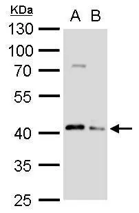
Figure 1. Western blot analysis of Apg3 using anti-Apg3 antibody (M01768). Electrophoresis was performed on a 5-20% SDS-PAGE gel at 70V (Stacking gel) / 90V (Resolving gel) for 2-3 hours. The sample well of each lane was loaded with 30 ug of sample under reducing conditions. Lane 1: human Jurkat whole cell lysates, Lane 2: human K562 whole cell lysates, Lane 3: human Raji whole cell lysates, Lane 4: monkey COS-7 whole cell lysates, Lane 5: rat C6 whole cell lysates, Lane 6: mouse kidney tissue lysates, Lane 7: mouse NIH/3T3 whole cell lysates. After electrophoresis, proteins were transferred to a nitrocellulose membrane at 150 mA for 50-90 minutes. Blocked the membrane with 5% non-fat milk/TBS for 1.5 hour at RT. The membrane was incubated with rabbit anti-Apg3 antigen affinity purified monoclonal antibody (Catalog # M01768) at 1:500 overnight at 4°C, then washed with TBS-0.1%Tween 3 times with 5 minutes each and probed with a goat anti-rabbit IgG-HRP secondary antibody at a dilution of 1:5000 for 1.5 hour at RT. The signal is developed using an Enhanced Chemiluminescent detection (ECL) kit (Catalog # EK1002) with Tanon 5200 system. A specific band was detected for Apg3 at approximately 40 kDa. The expected band size for Apg3 is at 36 kDa.
Figure 1. Western blot analysis of Apg3 using anti-Apg3 antibody (M01768). Electrophoresis was performed on a 5-20% SDS-PAGE gel at 70V (Stacking gel) / 90V (Resolving gel) for 2-3 hours. The sample well of each lane was loaded with 30 ug of sample under reducing conditions. Lane 1: human Jurkat whole cell lysates, Lane 2: human K562 whole cell lysates, Lane 3: human Raji whole cell lysates, Lane 4: monkey COS-7 whole cell lysates, Lane 5: rat C6 whole cell lysates, Lane 6: mouse kidney tissue lysates, Lane 7: mouse NIH/3T3 whole cell lysates. After electrophoresis, proteins were transferred to a nitrocellulose membrane at 150 mA for 50-90 minutes. Blocked the membrane with 5% non-fat milk/TBS for 1.5 hour at RT. The membrane was incubated with rabbit anti-Apg3 antigen affinity purified monoclonal antibody (Catalog # M01768) at 1:500 overnight at 4°C, then washed with TBS-0.1%Tween 3 times with 5 minutes each and probed with a goat anti-rabbit IgG-HRP secondary antibody at a dilution of 1:5000 for 1.5 hour at RT. The signal is developed using an Enhanced Chemiluminescent detection (ECL) kit (Catalog # EK1002) with Tanon 5200 system. A specific band was detected for Apg3 at approximately 40 kDa. The expected band size for Apg3 is at 36 kDa.
Anti-Apg3 (Atg3) Rabbit Monoclonal Antibody
M01768
ApplicationsImmunoFluorescence, Western Blot, ImmunoCytoChemistry, ImmunoHistoChemistry
Product group Antibodies
ReactivityHuman, Mouse, Rat
TargetATG3
Overview
- SupplierBoster Bio
- Product NameAnti-Apg3 (Atg3) Rabbit Monoclonal Antibody
- Delivery Days Customer9
- ApplicationsImmunoFluorescence, Western Blot, ImmunoCytoChemistry, ImmunoHistoChemistry
- CertificationResearch Use Only
- ClonalityMonoclonal
- Clone IDABBC-1
- Gene ID64422
- Target nameATG3
- Target descriptionautophagy related 3
- Target synonymsAPG3, APG3-LIKE, APG3L, PC3-96, hApg3, ubiquitin-like-conjugating enzyme ATG3, APG3 autophagy 3-like, ATG3 autophagy related 3 homolog, autophagy-related protein 3
- HostRabbit
- IsotypeIgG
- Protein IDQ9NT62
- Protein NameUbiquitin-like-conjugating enzyme ATG3
- Scientific DescriptionBoster Bio Anti-Apg3 (Atg3) Rabbit Monoclonal Antibody catalog # M01768. Tested in WB, IHC, ICC/IF applications. This antibody reacts with Human, Mouse, Rat.
- ReactivityHuman, Mouse, Rat
- Storage Instruction-20°C
- UNSPSC12352203
References
- Xing JJ, Hou JG, Ma ZN, et al. Ginsenoside Rb3 provides protective effects against cisplatin-induced nephrotoxicity via regulation of AMPK-/mTOR-mediated autophagy and inhibition of apoptosis in vitro and in vivo. Cell Prolif. 2019,52(4):e12627. doi: 10.1111/cpr.12627Read this paper
Datasheet
MSDS
Related products
Product group Antibodies
Anti-ATG3 Antibody144-05809
ApplicationsWestern Blot, ImmunoHistoChemistry
ReactivityHuman, Mouse
TargetATG3
- SizePrice

Product group Antibodies
Anti-Apg3/ATG3 Antibody Picoband(r)A01768-1-CARRIER-FREE
ApplicationsWestern Blot
ReactivityHuman, Monkey, Mouse
TargetATG3
- SizePrice
Product group Antibodies
ATG3 antibodyGTX128065
ApplicationsWestern Blot
ReactivityHuman, Mouse, Rat
TargetATG3
- SizePrice
Product group Antibodies
Anti-ATG3 AntibodyA31063
ApplicationsWestern Blot, ImmunoHistoChemistry
ReactivityHuman, Mouse, Rat
- SizePrice
Product group Antibodies
Atg3 Polyclonal AntibodyCAC07035
ApplicationsImmunoFluorescence, Western Blot, ELISA, ImmunoHistoChemistry
TargetATG3
- SizePrice
Product group Antibodies
ATG3 Recombinant Antibody, AbBy Fluor-488 ConjugatedBSM-61474R-BF488
ApplicationsImmunoFluorescence, Western Blot, ImmunoCytoChemistry
ReactivityHuman, Mouse, Rat
TargetATG3
- SizePrice
Product group Antibodies
ATG3 AntibodyCSB-PA002288HA01HU
ApplicationsImmunoFluorescence, Western Blot, ELISA, ImmunoHistoChemistry
ReactivityHuman
TargetATG3
- SizePrice
Product group Antibodies

ATG3 AntibodyLS-C401261
ApplicationsELISA, ImmunoHistoChemistry
ReactivityHuman, Mouse, Rat
TargetATG3
- SizePrice

Product group Antibodies
Anti-ATG3 AntibodyHPA040471
ApplicationsImmunoHistoChemistry
ReactivityHuman
TargetATG3
- SizePrice