


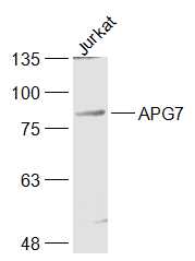
Figure 1. Western blot analysis of Apg7/ATG7 using anti-Apg7/ATG7 antibody (PA2261). Electrophoresis was performed on a 5-20% SDS-PAGE gel at 70V (Stacking gel) / 90V (Resolving gel) for 2-3 hours. The sample well of each lane was loaded with 30 ug of sample under reducing conditions. Lane 1: human Hela whole cell lysates, Lane 2: human HepG2 whole cell lysates, Lane 3: human K562 whole cell lysates, Lane 4: human Jurkat whole cell lysates, Lane 5: rat kidney tissue lysates, Lane 6: mouse kidney tissue lysates, Lane 7: mouse spleen tissue lysates, Lane 8: mouse SP2/0 whole cell lysates. After electrophoresis, proteins were transferred to a nitrocellulose membrane at 150 mA for 50-90 minutes. Blocked the membrane with 5% non-fat milk/TBS for 1.5 hour at RT. The membrane was incubated with rabbit anti-Apg7/ATG7 antigen affinity purified polyclonal antibody (Catalog # PA2261) at 0.5 microg/mL overnight at 4°C, then washed with TBS-0.1%Tween 3 times with 5 minutes each and probed with a goat anti-rabbit IgG-HRP secondary antibody at a dilution of 1:5000 for 1.5 hour at RT. The signal is developed using an Enhanced Chemiluminescent detection (ECL) kit (Catalog # EK1002) with Tanon 5200 system. A specific band was detected for Apg7/ATG7 at approximately 78 kDa. The expected band size for Apg7/ATG7 is at 78 kDa.
. Overlay histogram showing Hela cells stained with PA2261 (Blue line). To facilitate intracellular staining, cells were fixed with 4% paraformaldehyde and permeabilized with permeabilization buffer. The cells were blocked with 10% normal goat serum. And then incubated with rabbit anti-Apg7/ATG7 Antibody (PA2261, 1 microg/1x106 cells) for 30 min at 20°C. DyLight®488 conjugated goat anti-rabbit IgG (BA1127, 5-10 microg/1x106 cells) was used as secondary antibody for 30 minutes at 20°C. Isotype control antibody (Green line) was rabbit IgG (1 microg/1x106) used under the same conditions. Unlabelled sample without incubation with primary antibody and secondary antibody (Red line) was used as a blank control.")
Figure 1. Western blot analysis of Apg7/ATG7 using anti-Apg7/ATG7 antibody (PA2261). Electrophoresis was performed on a 5-20% SDS-PAGE gel at 70V (Stacking gel) / 90V (Resolving gel) for 2-3 hours. The sample well of each lane was loaded with 30 ug of sample under reducing conditions. Lane 1: human Hela whole cell lysates, Lane 2: human HepG2 whole cell lysates, Lane 3: human K562 whole cell lysates, Lane 4: human Jurkat whole cell lysates, Lane 5: rat kidney tissue lysates, Lane 6: mouse kidney tissue lysates, Lane 7: mouse spleen tissue lysates, Lane 8: mouse SP2/0 whole cell lysates. After electrophoresis, proteins were transferred to a nitrocellulose membrane at 150 mA for 50-90 minutes. Blocked the membrane with 5% non-fat milk/TBS for 1.5 hour at RT. The membrane was incubated with rabbit anti-Apg7/ATG7 antigen affinity purified polyclonal antibody (Catalog # PA2261) at 0.5 microg/mL overnight at 4°C, then washed with TBS-0.1%Tween 3 times with 5 minutes each and probed with a goat anti-rabbit IgG-HRP secondary antibody at a dilution of 1:5000 for 1.5 hour at RT. The signal is developed using an Enhanced Chemiluminescent detection (ECL) kit (Catalog # EK1002) with Tanon 5200 system. A specific band was detected for Apg7/ATG7 at approximately 78 kDa. The expected band size for Apg7/ATG7 is at 78 kDa.
Anti-Apg7/ATG7 Antibody Picoband(r)
PA2261
ApplicationsFlow Cytometry, Western Blot
Product group Antibodies
ReactivityChicken, Human, Mouse, Rat
TargetATG7
Overview
- SupplierBoster Bio
- Product NameAnti-Apg7/ATG7 Antibody Picoband(r)
- Delivery Days Customer9
- Application Supplier NoteTested Species: In-house tested species with positive results. Predicted Species: Species predicted to be fit for the product based on sequence similarities. Other applications have not been tested. Optimal dilutions should be determined by end users.
- ApplicationsFlow Cytometry, Western Blot
- Applications SupplierWB
- CertificationResearch Use Only
- ClonalityPolyclonal
- Concentration500 ug/ml
- Gene ID10533
- Target nameATG7
- Target descriptionautophagy related 7
- Target synonymsAPG7-LIKE, APG7L, GSA7, SCAR31, ubiquitin-like modifier-activating enzyme ATG7, APG7 autophagy 7-like, ATG12-activating enzyme E1 ATG7, hAGP7, ubiquitin-activating enzyme E1-like protein
- HostRabbit
- IsotypeIgG
- Protein IDO95352
- Protein NameUbiquitin-like modifier-activating enzyme ATG7
- Scientific DescriptionBoster Bio Anti-Apg7/ATG7 Antibody catalog # PA2261. Tested in Flow Cytometry, WB applications. This antibody reacts with Human, Mouse, Rat. The brand Picoband indicates this is a premium antibody that guarantees superior quality, high affinity, and strong signals with minimal background in Western blot applications. Only our best-performing antibodies are designated as Picoband, ensuring unmatched performance.
- ReactivityChicken, Human, Mouse, Rat
- Reactivity SupplierHuman, Mouse, Rat, Chicken
- Storage Instruction-20°C,2°C to 8°C
- UNSPSC12352203
Datasheet
MSDS
Related products
Product group Antibodies
Anti-ATG7 Antibody144-00691
ApplicationsWestern Blot, ImmunoHistoChemistry
ReactivityHuman, Mouse, Rat
TargetATG7
- SizePrice

Product group Antibodies
Anti-ATG7 Antibody Picoband(r)A00346-4-CARRIER-FREE
ApplicationsFlow Cytometry, Western Blot, ELISA
ReactivityHuman
TargetATG7
- SizePrice
![Whole cell extract (30 μg) was separated by 7.5% SDS-PAGE, and the membrane was blotted with ATG7 antibody [N3C2], Internal (GTX113613) diluted at 1:1000. The HRP-conjugated anti-rabbit IgG antibody (GTX213110-01) was used to detect the primary antibody, and the signal was developed with Trident ECL plus-Enhanced.](https://www.genetex.com/upload/website/prouct_img/normal/GTX113613/GTX113613_40142_20190913_WB_M_w_23060501_290.webp)
Product group Antibodies
References
ATG7 antibody [N3C2], InternalGTX113613
ApplicationsWestern Blot
ReactivityHuman, Mouse
TargetATG7
- SizePrice
Product group Antibodies
Atg7 Polyclonal AntibodyCAC08251
ApplicationsImmunoFluorescence, Western Blot, ELISA, ImmunoHistoChemistry
TargetATG7
- SizePrice
Product group Antibodies
References
ATG7/APG7 Polyclonal AntibodyBS-2432R
ApplicationsImmunoFluorescence, Western Blot, ELISA, ImmunoCytoChemistry, ImmunoHistoChemistry, ImmunoHistoChemistry Frozen, ImmunoHistoChemistry Paraffin
ReactivityBovine, Canine, Chicken, Equine, Human, Mouse, Porcine, Rat
TargetATG7
- SizePrice
Product group Antibodies
ATG7 Monoclonal AntibodyCSB-MA617582
ApplicationsELISA, ImmunoHistoChemistry
ReactivityHuman, Mouse, Rat
TargetATG7
- SizePrice
Product group Antibodies

Apg7 / ATG7 Antibody (clone 1A1)LS-C764703
ApplicationsImmunoFluorescence, ImmunoHistoChemistry, ImmunoHistoChemistry Paraffin
ReactivityHuman, Mouse, Rat
TargetATG7
- SizePrice

Product group Antibodies
Anti-ATG7 AntibodyHPA007639
ApplicationsImmunoCytoChemistry, ImmunoHistoChemistry
ReactivityHuman
TargetATG7
- SizePrice