


Anti-APH1A Antibody
A89261
ApplicationsImmunoFluorescence, Western Blot, ImmunoCytoChemistry
Product group Antibodies
ReactivityHuman, Mouse
Overview
- SupplierAntibodies.com
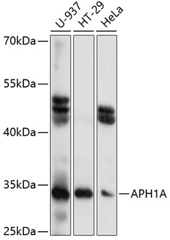
- Product NameAnti-APH1A Antibody
- Delivery Days Customer7
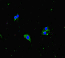
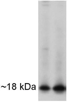
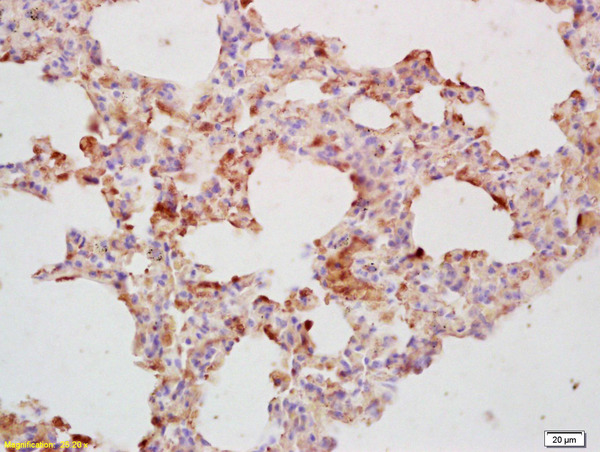
- ApplicationsImmunoFluorescence, Western Blot, ImmunoCytoChemistry
- CertificationResearch Use Only
- ClonalityPolyclonal
- ConjugateUnconjugated
- HostRabbit
- IsotypeIgG
- Scientific DescriptionRabbit polyclonal antibody to APH1A.
- ReactivityHuman, Mouse
- UNSPSC12352203
Related products
Product group Antibodies

APH1A / APH-1 AntibodyLS-C749656
ApplicationsWestern Blot
ReactivityHuman
TargetAPH1A
- SizePrice
Product group Antibodies

Goat anti-APH1AEB07860
ApplicationsWestern Blot, ELISA, ImmunoHistoChemistry
ReactivityBovine, Canine, Human, Mouse, Porcine, Rat
TargetAPH1A
- SizePrice

Product group Antibodies
Anti-APH1A Antibody Picoband(r)A04859-1-CARRIER-FREE
ApplicationsFlow Cytometry, ImmunoFluorescence, Western Blot, ImmunoHistoChemistry
ReactivityHuman, Mouse, Rat
TargetAPH1A
- SizePrice
Product group Antibodies
APH1A AntibodyCSB-PA853390LA01HU
ApplicationsImmunoFluorescence, ELISA
ReactivityHuman
TargetAPH1A
- SizePrice
Product group Antibodies
APH1A antibodyGTX79306
ApplicationsFlow Cytometry, ImmunoFluorescence, ImmunoPrecipitation, Western Blot, ImmunoCytoChemistry
ReactivityHamster, Human, Mouse
TargetAPH1A
- SizePrice
Product group Antibodies
Anti-APH1A Antibody144-60745
ApplicationsWestern Blot
ReactivityHuman
TargetAPH1A
- SizePrice
Product group Antibodies
APH1A Polyclonal AntibodyBS-4259R
ApplicationsFlow Cytometry, ImmunoFluorescence, Western Blot, ELISA, ImmunoCytoChemistry, ImmunoHistoChemistry, ImmunoHistoChemistry Frozen, ImmunoHistoChemistry Paraffin
ReactivityBovine, Canine, Equine, Human, Mouse, Porcine, Rabbit, Rat
TargetAPH1A
- SizePrice