

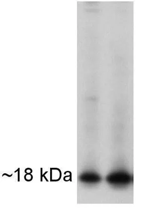
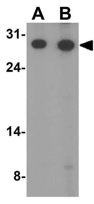
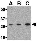
WB analysis of mouse brain and kidney extracts using GTX79306 APH1A antibody.


WB analysis of mouse brain and kidney extracts using GTX79306 APH1A antibody.
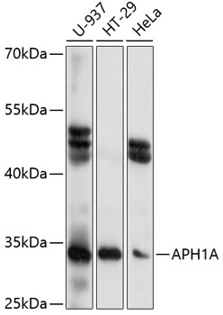
APH1A antibody
GTX79306
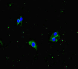
ApplicationsFlow Cytometry, ImmunoFluorescence, ImmunoPrecipitation, Western Blot, ImmunoCytoChemistry
Product group Antibodies
ReactivityHamster, Human, Mouse
TargetAPH1A
Overview
- SupplierGeneTex
- Product NameAPH1A antibody
- Delivery Days Customer9
- Application Supplier NoteWB: 1:1000. ICC/IF: 2 microg/mL. FCM: 3-5 microg/106 cells. *Optimal dilutions/concentrations should be determined by the researcher.Not tested in other applications.
- ApplicationsFlow Cytometry, ImmunoFluorescence, ImmunoPrecipitation, Western Blot, ImmunoCytoChemistry
- CertificationResearch Use Only
- ClonalityPolyclonal
- Concentration0.69 mg/ml
- ConjugateUnconjugated
- Gene ID51107
- Target nameAPH1A
- Target descriptionaph-1A gamma-secretase subunit
- Target synonyms6530402N02Rik, APH-1, APH-1A, CGI-78, gamma-secretase subunit APH-1A, APH1A gamma secretase subunit, anterior pharynx defective 1 homolog A, aph-1 homolog A, gamma-secretase subunit, aph-1alpha, presenilin-stabilization factor
- HostRabbit
- IsotypeIgG
- Protein IDQ96BI3
- Protein NameGamma-secretase subunit APH-1A
- Scientific DescriptionThis gene encodes a component of the gamma secretase complex that cleaves integral membrane proteins such as Notch receptors and beta-amyloid precursor protein. The gamma secretase complex contains this gene product, or the paralogous anterior pharynx defective 1 homolog B (APH1B), along with the presenilin, nicastrin, and presenilin enhancer-2 proteins. The precise function of this seven-transmembrane-domain protein is unknown though it is suspected of facilitating the association of nicastrin and presenilin in the gamma secretase complex as well as interacting with substrates of the gamma secretase complex prior to their proteolytic processing. Polymorphisms in a promoter region of this gene have been associated with an increased risk for developing sporadic Alzheimers disease. Alternative splicing results in multiple protein-coding and non-protein-coding transcript variants. [provided by RefSeq, Aug 2011]
- ReactivityHamster, Human, Mouse
- Storage Instruction-20°C or -80°C,2°C to 8°C
- UNSPSC41116161
Datasheet
Related products
Product group Antibodies
Anti-APH1A AntibodyA89261
ApplicationsImmunoFluorescence, Western Blot, ImmunoCytoChemistry
ReactivityHuman, Mouse
- SizePrice
Product group Antibodies

APH1A / APH-1 AntibodyLS-C749656
ApplicationsWestern Blot
ReactivityHuman
TargetAPH1A
- SizePrice
Product group Antibodies

Goat anti-APH1AEB07860
ApplicationsWestern Blot, ELISA, ImmunoHistoChemistry
ReactivityBovine, Canine, Human, Mouse, Porcine, Rat
TargetAPH1A
- SizePrice

Product group Antibodies
Anti-APH1A Antibody Picoband(r)A04859-1-CARRIER-FREE
ApplicationsFlow Cytometry, ImmunoFluorescence, Western Blot, ImmunoHistoChemistry
ReactivityHuman, Mouse, Rat
TargetAPH1A
- SizePrice
Product group Antibodies
APH1A AntibodyCSB-PA853390LA01HU
ApplicationsImmunoFluorescence, ELISA
ReactivityHuman
TargetAPH1A
- SizePrice
Product group Antibodies
APH1A antibodyGTX85061
ApplicationsWestern Blot, ELISA, ImmunoHistoChemistry, ImmunoHistoChemistry Paraffin
ReactivityHuman, Mouse, Rat
TargetAPH1A
- SizePrice
Product group Antibodies
APH1A antibodyGTX85062
ApplicationsWestern Blot, ELISA
ReactivityHuman, Mouse
TargetAPH1A
- SizePrice
Product group Antibodies
APH1A antibody, InternalGTX89025
ApplicationsWestern Blot, ImmunoHistoChemistry, ImmunoHistoChemistry Paraffin
ReactivityHuman
TargetAPH1A
- SizePrice
Product group Antibodies
Anti-APH1A Antibody144-60745
ApplicationsWestern Blot
ReactivityHuman
TargetAPH1A
- SizePrice
Product group Antibodies
APH1A Polyclonal AntibodyBS-4259R
ApplicationsFlow Cytometry, ImmunoFluorescence, Western Blot, ELISA, ImmunoCytoChemistry, ImmunoHistoChemistry, ImmunoHistoChemistry Frozen, ImmunoHistoChemistry Paraffin
ReactivityBovine, Canine, Equine, Human, Mouse, Porcine, Rabbit, Rat
TargetAPH1A
- SizePrice