


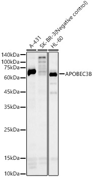
Figure 1. Western blot analysis of APOBEC3B using anti-APOBEC3B antibody (A01088-1). Electrophoresis was performed on a 5-20% SDS-PAGE gel at 70V (Stacking gel) / 90V (Resolving gel) for 2-3 hours. The sample well of each lane was loaded with 30 ug of sample under reducing conditions. Lane 1: human HepG2 whole cell lysates, Lane 2: human Jurkat whole cell lysates, Lane 3: human THP-1 whole cell lysates, Lane 4: human HEK293 whole cell lysates. After electrophoresis, proteins were transferred to a nitrocellulose membrane at 150 mA for 50-90 minutes. Blocked the membrane with 5% non-fat milk/TBS for 1.5 hour at RT. The membrane was incubated with rabbit anti-APOBEC3B antigen affinity purified polyclonal antibody (Catalog # A01088-1) at 0.5 microg/mL overnight at 4°C, then washed with TBS-0.1%Tween 3 times with 5 minutes each and probed with a goat anti-rabbit IgG-HRP secondary antibody at a dilution of 1:5000 for 1.5 hour at RT. The signal is developed using an Enhanced Chemiluminescent detection (ECL) kit (Catalog # EK1002) with Tanon 5200 system. A specific band was detected for APOBEC3B at approximately 35 kDa. The expected band size for APOBEC3B is at 46 kDa.
Figure 1. Western blot analysis of APOBEC3B using anti-APOBEC3B antibody (A01088-1). Electrophoresis was performed on a 5-20% SDS-PAGE gel at 70V (Stacking gel) / 90V (Resolving gel) for 2-3 hours. The sample well of each lane was loaded with 30 ug of sample under reducing conditions. Lane 1: human HepG2 whole cell lysates, Lane 2: human Jurkat whole cell lysates, Lane 3: human THP-1 whole cell lysates, Lane 4: human HEK293 whole cell lysates. After electrophoresis, proteins were transferred to a nitrocellulose membrane at 150 mA for 50-90 minutes. Blocked the membrane with 5% non-fat milk/TBS for 1.5 hour at RT. The membrane was incubated with rabbit anti-APOBEC3B antigen affinity purified polyclonal antibody (Catalog # A01088-1) at 0.5 microg/mL overnight at 4°C, then washed with TBS-0.1%Tween 3 times with 5 minutes each and probed with a goat anti-rabbit IgG-HRP secondary antibody at a dilution of 1:5000 for 1.5 hour at RT. The signal is developed using an Enhanced Chemiluminescent detection (ECL) kit (Catalog # EK1002) with Tanon 5200 system. A specific band was detected for APOBEC3B at approximately 35 kDa. The expected band size for APOBEC3B is at 46 kDa.
Anti-APOBEC3B Antibody Picoband(r)
A01088-1-CARRIER-FREE
ApplicationsWestern Blot
Product group Antibodies
ReactivityHuman
TargetAPOBEC3B
Overview
- SupplierBoster Bio
- Product NameAnti-APOBEC3B Antibody Picoband(r)
- Delivery Days Customer9
- ApplicationsWestern Blot
- CertificationResearch Use Only
- ClonalityPolyclonal
- Concentration500 ug/ml
- Gene ID9582
- Target nameAPOBEC3B
- Target descriptionapolipoprotein B mRNA editing enzyme catalytic subunit 3B
- Target synonymsA3B, APOBEC1L, ARCD3, ARP4, DJ742C19.2, PHRBNL, bK150C2.2, DNA dC->dU-editing enzyme APOBEC-3B, apolipoprotein B mRNA editing enzyme, catalytic polypeptide-like 3B, cytidine deaminase, phorbolin 2, phorbolin 3, phorbolin-1-related protein, phorbolin-2/3, probable DNA dC->dU-editing enzyme APOBEC-3B
- HostRabbit
- IsotypeIgG
- Protein IDQ9UH17
- Protein NameDNA dC->dU-editing enzyme APOBEC-3B
- Scientific DescriptionBoster Bio Anti-APOBEC3B Antibody Picoband® catalog # A01088-1. Tested in WB applications. This antibody reacts with HumanThe brand Picoband indicates this is a premium antibody that guarantees superior quality, high affinity, and strong signals with minimal background in Western blot applications. Only our best-performing antibodies are designated as Picoband, ensuring unmatched performance.
- ReactivityHuman
- Storage Instruction-20°C,2°C to 8°C
- UNSPSC12352203
Related products
Product group Antibodies
Anti-APOBEC3B AntibodyA10590
ApplicationsWestern Blot
ReactivityHuman
- SizePrice

Product group Antibodies
Anti-APOBEC3B AntibodyHPA066719
ApplicationsImmunoCytoChemistry
ReactivityHuman
TargetAPOBEC3B
- SizePrice
Product group Antibodies

APOBEC3B AntibodyLS-C410544
ApplicationsWestern Blot
ReactivityHuman
TargetAPOBEC3B
- SizePrice
Product group Antibodies
References
APOBEC3B antibodyGTX17214
ApplicationsWestern Blot, ELISA, ImmunoHistoChemistry, ImmunoHistoChemistry Paraffin
ReactivityHuman, Mouse, Rat
TargetAPOBEC3B
- SizePrice
Product group Antibodies
Anti-APOBEC3B Antibody144-09010
ApplicationsWestern Blot
ReactivityHuman
TargetAPOBEC3B
- SizePrice