


Figure 1. Western blot analysis of AQP3 using anti-AQP3 antibody (PA1488). Electrophoresis was performed on a 5-20% SDS-PAGE gel at 70V (Stacking gel) / 90V (Resolving gel) for 2-3 hours. The sample well of each lane was loaded with 30 ug of sample under reducing conditions. Lane 1: rat kidney tissue lysates, Lane 2: rat kidney tissue lysates. Lane 3: mouse kidney tissue lysates. Lane 4: mouse kidney tissue lysates. After electrophoresis, proteins were transferred to a nitrocellulose membrane at 150 mA for 50-90 minutes. Blocked the membrane with 5% non-fat milk/TBS for 1.5 hour at RT. The membrane was incubated with rabbit anti-AQP3 antigen affinity purified polyclonal antibody (Catalog # PA1488) at 0.5 microg/mL overnight at 4°C, then washed with TBS-0.1%Tween 3 times with 5 minutes each and probed with a goat anti-rabbit IgG-HRP secondary antibody at a dilution of 1:5000 for 1.5 hour at RT. The signal is developed using an Enhanced Chemiluminescent detection (ECL) kit (Catalog # EK1002) with Tanon 5200 system. A specific band was detected for AQP3 at approximately 32-36 kDa. The expected band size for AQP3 is at 32 kDa.
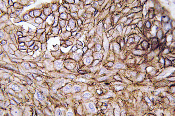
. AQP3 was detected in a paraffin-embedded section of human bladder cancer tissue. Heat mediated antigen retrieval was performed in EDTA buffer (pH 8.0, epitope retrieval solution). The tissue section was blocked with 10% goat serum. The tissue section was then incubated with 2 microg/ml rabbit anti-AQP3 Antibody (PA1488) overnight at 4°C. Peroxidase Conjugated Goat Anti-rabbit IgG was used as secondary antibody and incubated for 30 minutes at 37°C. The tissue section was developed using HRP Conjugated Rabbit IgG Super Vision Assay Kit (Catalog # SV0002) with DAB as the chromogen.")
. AQP3 was detected in a paraffin-embedded section of human cervical cancer tissue. Heat mediated antigen retrieval was performed in EDTA buffer (pH 8.0, epitope retrieval solution). The tissue section was blocked with 10% goat serum. The tissue section was then incubated with 2 microg/ml rabbit anti-AQP3 Antibody (PA1488) overnight at 4°C. Peroxidase Conjugated Goat Anti-rabbit IgG was used as secondary antibody and incubated for 30 minutes at 37°C. The tissue section was developed using HRP Conjugated Rabbit IgG Super Vision Assay Kit (Catalog # SV0002) with DAB as the chromogen.")
. AQP3 was detected in a paraffin-embedded section of mouse kidney tissue. Heat mediated antigen retrieval was performed in EDTA buffer (pH 8.0, epitope retrieval solution). The tissue section was blocked with 10% goat serum. The tissue section was then incubated with 2 microg/ml rabbit anti-AQP3 Antibody (PA1488) overnight at 4°C. Peroxidase Conjugated Goat Anti-rabbit IgG was used as secondary antibody and incubated for 30 minutes at 37°C. The tissue section was developed using HRP Conjugated Rabbit IgG Super Vision Assay Kit (Catalog # SV0002) with DAB as the chromogen.")
. AQP3 was detected in a paraffin-embedded section of rat kidney tissue. Heat mediated antigen retrieval was performed in EDTA buffer (pH 8.0, epitope retrieval solution). The tissue section was blocked with 10% goat serum. The tissue section was then incubated with 2 microg/ml rabbit anti-AQP3 Antibody (PA1488) overnight at 4°C. Peroxidase Conjugated Goat Anti-rabbit IgG was used as secondary antibody and incubated for 30 minutes at 37°C. The tissue section was developed using HRP Conjugated Rabbit IgG Super Vision Assay Kit (Catalog # SV0002) with DAB as the chromogen.")
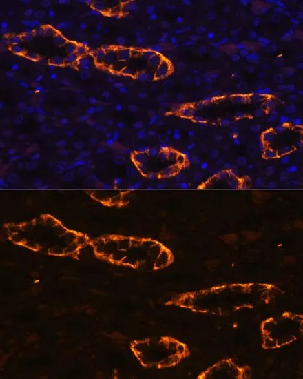
. AQP3 was detected in immunocytochemical section of Hela cells. Enzyme antigen retrieval was performed using IHC enzyme antigen retrieval reagent (AR0022) for 15 mins. The cells were blocked with 10% goat serum. And then incubated with 5microg/mL rabbit anti- AQP3 Antibody (PA1488) overnight at 4°C. Cy3 Conjugated Goat Anti-Rabbit IgG (BA1032) was used as secondary antibody at 1:500 dilution and incubated for 30 minutes at 37°C. The section was counterstained with DAPI. Visualize using a fluorescence microscope and filter sets appropriate for the label used.")
. Overlay histogram showing RT4 cells stained with PA1488 (Blue line). The cells were fixed with 4% paraformaldehyde and blocked with 10% normal goat serum. And then incubated with rabbit anti-AQP3 Antibody (PA1488, 1 microg/1x106 cells) for 30 min at 20°C. DyLight®488 conjugated goat anti-rabbit IgG (BA1127, 5-10 microg/1x106 cells) was used as secondary antibody for 30 minutes at 20°C. Isotype control antibody (Green line) was rabbit IgG (1 microg/1x106) used under the same conditions. Unlabelled sample without incubation with primary antibody and secondary antibody (Red line) was used as a blank control.")
Figure 1. Western blot analysis of AQP3 using anti-AQP3 antibody (PA1488). Electrophoresis was performed on a 5-20% SDS-PAGE gel at 70V (Stacking gel) / 90V (Resolving gel) for 2-3 hours. The sample well of each lane was loaded with 30 ug of sample under reducing conditions. Lane 1: rat kidney tissue lysates, Lane 2: rat kidney tissue lysates. Lane 3: mouse kidney tissue lysates. Lane 4: mouse kidney tissue lysates. After electrophoresis, proteins were transferred to a nitrocellulose membrane at 150 mA for 50-90 minutes. Blocked the membrane with 5% non-fat milk/TBS for 1.5 hour at RT. The membrane was incubated with rabbit anti-AQP3 antigen affinity purified polyclonal antibody (Catalog # PA1488) at 0.5 microg/mL overnight at 4°C, then washed with TBS-0.1%Tween 3 times with 5 minutes each and probed with a goat anti-rabbit IgG-HRP secondary antibody at a dilution of 1:5000 for 1.5 hour at RT. The signal is developed using an Enhanced Chemiluminescent detection (ECL) kit (Catalog # EK1002) with Tanon 5200 system. A specific band was detected for AQP3 at approximately 32-36 kDa. The expected band size for AQP3 is at 32 kDa.
Anti-Aquaporin 3/AQP3 Antibody Picoband(r)
PA1488
ApplicationsFlow Cytometry, ImmunoFluorescence, Western Blot, ImmunoCytoChemistry, ImmunoHistoChemistry
Product group Antibodies
ReactivityBovine, Human, Mouse, Rat
TargetAQP3
Overview
- SupplierBoster Bio
- Product NameAnti-Aquaporin 3/AQP3 Antibody Picoband(r)
- Delivery Days Customer9
- Application Supplier NoteTested Species: In-house tested species with positive results. By Heat: Boiling the paraffin sections in 10mM citrate buffer, pH6.0, for 20mins is required for the staining of formalin/paraffin sections. Other applications have not been tested. Optimal dilutions should be determined by end users.
- ApplicationsFlow Cytometry, ImmunoFluorescence, Western Blot, ImmunoCytoChemistry, ImmunoHistoChemistry
- Applications SupplierIHP, IHF, ICC, WB, IHC
- CertificationResearch Use Only
- ClonalityPolyclonal
- Concentration500 ug/ml
- Gene ID360
- Target nameAQP3
- Target descriptionaquaporin 3 (Gill blood group)
- Target synonymsAQP-3, GIL, aquaporin-3, aquaglyceroporin-3, aquaporin 3 (GIL blood group)
- HostRabbit
- IsotypeIgG
- Protein IDQ92482
- Protein NameAquaporin-3
- Scientific DescriptionBoster Bio Anti-Aquaporin 3/AQP3 Antibody catalog # PA1488. Tested in Flow Cytometry, IF, IHC, ICC, WB applications. This antibody reacts with Human, Mouse, Rat. The brand Picoband indicates this is a premium antibody that guarantees superior quality, high affinity, and strong signals with minimal background in Western blot applications. Only our best-performing antibodies are designated as Picoband, ensuring unmatched performance.
- ReactivityBovine, Human, Mouse, Rat
- Reactivity SupplierHuman, Mouse, Rat, Bovine
- Storage Instruction-20°C,2°C to 8°C
- UNSPSC12352203
References
- Deng Y, Zhao Q, Zhou HY, et al. Activation of ASIC3/ERK pathway by paeoniflorin improves intestinal fluid metabolism and visceral sensitivity in slow transit constipated rats. Kaohsiung J Med Sci. 2024,40(6):561-574. doi: 10.1002/kjm2.12829Read this paper
- Lv H, Niu J, Pan W, et al. Stool-softening effect and action mechanism of free anthraquinones extracted from Rheum palmatum L. on water deficit-induced constipation in rats. J Ethnopharmacol. 2024,319(Pt 3):117336. doi: 10.1016/j.jep.2023.117336Read this paper
- Pan S, Lan Y, Chen B, et al. Tanshinone IIA changed the amniotic fluid volume and regulated expression of AQP1 and AQP3 in amniotic epithelium cells: a promising drug treating abnormal amniotic fluid volume. Mol Med. 2023,29(1):83. doi: 10.1186/s10020-023-00687-6Read this paper
- Pellavio G, Sommi P, Anselmi-Tamburini U, et al. Cerium Oxide Nanoparticles Regulate Oxidative Stress in HeLa Cells by Increasing the Aquaporin-Mediated Hydrogen Peroxide Permeability. Int J Mol Sci. 2022,23(18). doi: 10.3390/ijms231810837Read this paper
- Shao H, Pan S, Lan Y, et al. Tanshinone IIA increased amniotic fluid volume through down-regulating placental AQPs expression via inhibiting the activity of GSK-3β. Cell Tissue Res. 2022,389(3):547-558. doi: 10.1007/s00441-022-03646-5Read this paper
- Chen X, Zhang X, Du M, et al. In vivo preclinical PET/CT imaging of carbon-11-labeled aminoglycerol probe for the diagnosis of liver fibrosis. Ann Nucl Med. 2019,33(11):806-812. doi: 10.1007/s12149-019-01391-4Read this paper
- Feng J, Yan S, Chen Y, et al. Aquaporin1-3 expression in normal and hydronephrotic kidneys in the human fetus. Pediatr Res. 2019,86(5):595-602. doi: 10.1038/s41390-019-0485-6Read this paper
- He J, Zeng L, Wei R, et al. Lagopsis supina exerts its diuretic effect via inhibition of aquaporin-1, 2 and 3 expression in a rat model of traumatic blood stasis. J Ethnopharmacol. 2019,231:446-452. doi: 10.1016/j.jep.2018.10.034Read this paper
- Shin SY, Lee DH, Gil HN, et al. Agerarin, identified from Ageratum houstonianum, stimulates circadian CLOCK-mediated aquaporin-3 gene expression in HaCaT keratinocytes. Sci Rep. 2017,7(1):11175. doi: 10.1038/s41598-017-11642-xRead this paper
- Li H, Zhou L, Dai J. Retinoic acid receptor-related orphan receptor RORα regulates differentiation and survival of keratinocytes during hypoxia. J Cell Physiol. 2018,233(1):641-650. doi: 10.1002/jcp.25924Read this paper
Datasheet
MSDS
Related products
Product group Antibodies
Anti-AQP3 AntibodyA97793
ApplicationsELISA, ImmunoHistoChemistry
ReactivityHuman, Mouse, Rat
- SizePrice
Product group Antibodies

AQP3 / Aquaporin 3 AntibodyLS-C764594
ApplicationsWestern Blot, ELISA, ImmunoHistoChemistry
ReactivityHuman, Mouse, Rat
TargetAQP3
- SizePrice

Product group Antibodies
Anti-AQP3 AntibodyHPA014924
ApplicationsImmunoCytoChemistry, ImmunoHistoChemistry
ReactivityHuman
TargetAQP3
- SizePrice
Product group Antibodies
AQP3 AntibodyCSB-PA009603
ApplicationsELISA, ImmunoHistoChemistry
ReactivityHuman, Mouse, Rat
TargetAQP3
- SizePrice
Product group Antibodies
Aqp3 Polyclonal AntibodyCAC10970
ApplicationsImmunoFluorescence, ELISA, ImmunoHistoChemistry
TargetAQP3
- SizePrice
Product group Antibodies
Anti-Aquaporin 3/AQP3 Antibody Picoband(r)PA1488-CARRIER-FREE
ApplicationsFlow Cytometry, ImmunoFluorescence, Western Blot, ImmunoCytoChemistry, ImmunoHistoChemistry
ReactivityBovine, Human, Mouse, Rat
TargetAQP3
- SizePrice
Product group Antibodies
Aquaporin 3 antibodyGTX64481
ApplicationsWestern Blot, ImmunoHistoChemistry, ImmunoHistoChemistry Paraffin
ReactivityHuman, Mouse, Rat
TargetAQP3
- SizePrice
Product group Antibodies
Anti-AQP3 Antibody144-02838
ApplicationsImmunoFluorescence, Western Blot, ImmunoHistoChemistry
ReactivityHuman, Mouse, Rat
TargetAQP3
- SizePrice