


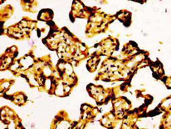
Immunohistochemical staining of human colorectal cancer shows weak to moderate nuclear positivity in tumor cells.

Immunohistochemical staining of human colorectal cancer shows weak to moderate nuclear positivity in tumor cells.
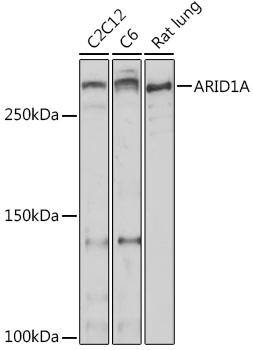
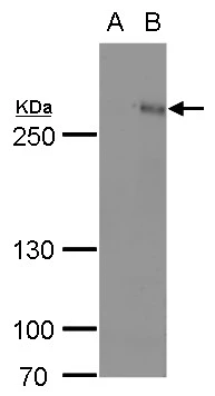
Anti-ARID1A Antibody
HPA005456
ApplicationsWestern Blot, ImmunoCytoChemistry, ImmunoHistoChemistry
Product group Antibodies
ReactivityHuman
TargetARID1A
Overview
- SupplierAtlas Antibodies
- Product NameAnti-ARID1A Antibody
- Delivery Days Customer4
- ApplicationsWestern Blot, ImmunoCytoChemistry, ImmunoHistoChemistry
- CertificationResearch Use Only
- ClonalityPolyclonal
- ConjugateUnconjugated
- Gene ID8289
- Target nameARID1A
- Target descriptionAT-rich interaction domain 1A
- Target synonymsB120, BAF250, BAF250a, BM029, C1orf4, CSS2, ELD, MRD14, OSA1, P270, SMARCF1, hELD, hOSA1, AT-rich interactive domain-containing protein 1A, ARID domain-containing protein 1A, AT rich interactive domain 1A (SWI-like), BRG1-associated factor 250a, OSA1 nuclear protein, SWI-like protein, SWI/SNF complex protein p270, SWI/SNF-related, matrix-associated, actin-dependent regulator of chromatin subfamily F member 1, brain protein 120, chromatin remodeling factor p250, osa homolog 1
- HostRabbit
- IsotypeIgG
- Protein IDO14497
- Protein NameAT-rich interactive domain-containing protein 1A
- Scientific DescriptionRecombinant Protein Epitope Signature Tag (PrEST) antigen sequence
- ReactivityHuman
- Storage Instruction-20°C,2°C to 8°C
- UNSPSC41116161
Datasheet
MSDS
Related products
Product group Antibodies
Anti-ARID1A AntibodyA306631
ApplicationsWestern Blot
ReactivityHuman, Mouse, Rat
- SizePrice
Product group Antibodies
Anti-ARID1A (C-term) Antibody102-24767
ApplicationsWestern Blot
TargetARID1A
- SizePrice

Product group Antibodies
Anti-ARID1A AntibodyAMAB91192
ApplicationsWestern Blot, ImmunoCytoChemistry, ImmunoHistoChemistry
ReactivityHuman
TargetARID1A
- SizePrice
Product group Antibodies
Anti-ARID1A AntibodyAMAB91192
ApplicationsWestern Blot, ImmunoCytoChemistry, ImmunoHistoChemistry
ReactivityHuman
TargetARID1A
- SizePrice

Product group Antibodies
Anti-ARID1A Antibody Picoband(r)A00247-2-CARRIER-FREE
ApplicationsFlow Cytometry, ImmunoFluorescence, Western Blot, ELISA, ImmunoCytoChemistry, ImmunoHistoChemistry
ReactivityHuman, Rat
TargetARID1A
- SizePrice
Product group Antibodies
ARID1A Recombinant Antibody, AbBy Fluor-350 ConjugatedBSM-61359R-BF350
ApplicationsImmunoFluorescence, Western Blot
ReactivityHuman, Mouse, Rat
TargetARID1A
- SizePrice
Product group Antibodies
ARID1A AntibodyCSB-PA002058LA01HU
ApplicationsELISA, ImmunoHistoChemistry
ReactivityHuman
TargetARID1A
- SizePrice
Product group Antibodies

ARID1A / BAF250 Antibody (aa95-108)LS-C402901
ApplicationsELISA, ImmunoHistoChemistry
ReactivityHuman, Mouse
TargetARID1A
- SizePrice
Product group Antibodies
ARID1A antibodyGTX129432
ApplicationsWestern Blot
ReactivityHuman
TargetARID1A
- SizePrice