


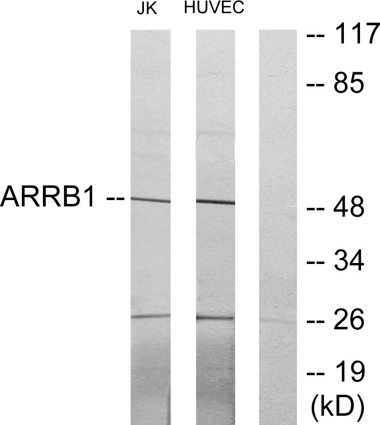
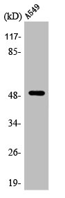
Figure 1. Western blot analysis of ARRB1 using anti-ARRB1 antibody (M02185). Electrophoresis was performed on a 5-20% SDS-PAGE gel at 70V (Stacking gel) / 90V (Resolving gel) for 2-3 hours. The sample well of each lane was loaded with 30 ug of sample under reducing conditions. Lane 1: human Jurkat whole cell lysates, Lane 2: human MCF-7 whole cell lysates, Lane 3: human K562 whoel cell lysates, Lane 4: rat spleen tissue lysates, Lane 5: rat PC-12 whoel cell lysates, Lane 6: mouse EL-4 whoel cell lysates. After electrophoresis, proteins were transferred to a nitrocellulose membrane at 150 mA for 50-90 minutes. Blocked the membrane with 5% non-fat milk/TBS for 1.5 hour at RT. The membrane was incubated with rabbit anti-ARRB1 antigen affinity purified monoclonal antibody (M02185) at 1:500 overnight at 4°C, then washed with TBS-0.1%Tween 3 times with 5 minutes each and probed with a goat anti-rabbit IgG-HRP secondary antibody at a dilution of 1:500 for 1.5 hour at RT. The signal is developed using an Enhanced Chemiluminescent detection (ECL) kit (Catalog # EK1002) with Tanon 5200 system. A specific band was detected for ARRB1 at approximately 50 kDa. The expected band size for ARRB1 is at 47 kDa.

Figure 1. Western blot analysis of ARRB1 using anti-ARRB1 antibody (M02185). Electrophoresis was performed on a 5-20% SDS-PAGE gel at 70V (Stacking gel) / 90V (Resolving gel) for 2-3 hours. The sample well of each lane was loaded with 30 ug of sample under reducing conditions. Lane 1: human Jurkat whole cell lysates, Lane 2: human MCF-7 whole cell lysates, Lane 3: human K562 whoel cell lysates, Lane 4: rat spleen tissue lysates, Lane 5: rat PC-12 whoel cell lysates, Lane 6: mouse EL-4 whoel cell lysates. After electrophoresis, proteins were transferred to a nitrocellulose membrane at 150 mA for 50-90 minutes. Blocked the membrane with 5% non-fat milk/TBS for 1.5 hour at RT. The membrane was incubated with rabbit anti-ARRB1 antigen affinity purified monoclonal antibody (M02185) at 1:500 overnight at 4°C, then washed with TBS-0.1%Tween 3 times with 5 minutes each and probed with a goat anti-rabbit IgG-HRP secondary antibody at a dilution of 1:500 for 1.5 hour at RT. The signal is developed using an Enhanced Chemiluminescent detection (ECL) kit (Catalog # EK1002) with Tanon 5200 system. A specific band was detected for ARRB1 at approximately 50 kDa. The expected band size for ARRB1 is at 47 kDa.
Anti-ARRB1/Beta Arrestin 1 Rabbit Monoclonal Antibody
M02185
ApplicationsFlow Cytometry, ImmunoFluorescence, Western Blot, ImmunoCytoChemistry, ImmunoHistoChemistry
Product group Antibodies
ReactivityHuman, Mouse, Rat
TargetARRB1
Overview
- SupplierBoster Bio
- Product NameAnti-ARRB1/Beta Arrestin 1 Rabbit Monoclonal Antibody
- Delivery Days Customer9
- ApplicationsFlow Cytometry, ImmunoFluorescence, Western Blot, ImmunoCytoChemistry, ImmunoHistoChemistry
- CertificationResearch Use Only
- ClonalityMonoclonal
- Clone IDCCB-1
- Gene ID408
- Target nameARRB1
- Target descriptionarrestin beta 1
- Target synonymsARB1, ARR1, beta-arrestin-1, arrestin 2, non-visual arrestin-2
- HostRabbit
- IsotypeIgG
- Protein IDP49407
- Protein NameBeta-arrestin-1
- Scientific DescriptionBoster Bio Anti-ARRB1/Beta Arrestin 1 Rabbit Monoclonal Antibody catalog # M02185. Tested in WB, IHC, ICC/IF, Flow Cytometry applications. This antibody reacts with Human, Mouse, Rat.
- ReactivityHuman, Mouse, Rat
- Storage Instruction-20°C
- UNSPSC12352203
Datasheet
MSDS
Related products
Product group Antibodies
Anti-ARRB1 AntibodyA97703
ApplicationsWestern Blot, ELISA
ReactivityHuman, Mouse, Rat
- SizePrice
Product group Antibodies
Anti-ARRB1 Antibody144-00998
ApplicationsImmunoFluorescence, ImmunoPrecipitation, Western Blot, ImmunoHistoChemistry
ReactivityHuman, Mouse, Rat
TargetARRB1
- SizePrice
Product group Antibodies
beta Arrestin 1 Recombinant AntibodyBSM-61185R
ApplicationsFlow Cytometry, ImmunoFluorescence, Western Blot, ImmunoCytoChemistry, ImmunoHistoChemistry, ImmunoHistoChemistry Frozen, ImmunoHistoChemistry Paraffin
TargetARRB1
- SizePrice
Product group Antibodies
ARRB1 AntibodyCSB-PA000935
ApplicationsImmunoFluorescence, Western Blot, ELISA, ImmunoHistoChemistry
ReactivityHuman, Monkey
TargetARRB1
- SizePrice
Product group Antibodies
Arrb1 Polyclonal AntibodyCAC11753
ApplicationsImmunoFluorescence, Western Blot, ELISA, ImmunoHistoChemistry
TargetARRB1
- SizePrice
![WB analysis of various samples using GTX02840 beta Arrestin 1 antibody [GT1243]. Dilution : 1:1000 Loading : 25μg](https://www.genetex.com/upload/website/prouct_img/normal/GTX02840/CutImage_A10742_WB_01_(1076419)_w_23053123_515.webp)
Product group Antibodies
ApplicationsWestern Blot
ReactivityHuman, Mouse
TargetARRB1
- SizePrice

Product group Antibodies
Anti-ARRB1 AntibodyHPA049318
ApplicationsImmunoCytoChemistry
ReactivityHuman
TargetARRB1
- SizePrice
Product group Antibodies

ARRB1 / Beta Arrestin 1 AntibodyLS-C400591
ApplicationsELISA, ImmunoHistoChemistry
ReactivityHuman, Mouse, Rat
TargetARRB1
- SizePrice