


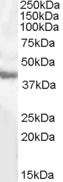
Figure 1. Western blot analysis of Aspartate Aminotransferase/GOT1 using anti-Aspartate Aminotransferase/GOT1 antibody (A04085-3). Electrophoresis was performed on a 5-20% SDS-PAGE gel at 70V (Stacking gel) / 90V (Resolving gel) for 2-3 hours. The sample well of each lane was loaded with 30ug of sample under reducing conditions. Lane 1: human placenta tissue lysates, Lane 2: human T-47D whole cell lysates, Lane 3: human HepG2 whole cell lysates, Lane 4: human Caco-2 whole cell lysates, Lane 5: human HL-60 whole cell lysates, Lane 6: human K562 whole cell lysates, Lane 7: human Hela whole cell lysates, Lane 8: rat brain tissue lysates, Lane 9: mouse brain tissue lysates, Lane 10: mouse liver tissue lysates. After Electrophoresis, proteins were transferred to a Nitrocellulose membrane at 150mA for 50-90 minutes. Blocked the membrane with 5% Non-fat Milk/ TBS for 1.5 hour at RT. The membrane was incubated with rabbit anti-Aspartate Aminotransferase/GOT1 antigen affinity purified polyclonal antibody (Catalog # A04085-3) at 0.25 microg/mL overnight at 4°C, then washed with TBS-0.1%Tween 3 times with 5 minutes each and probed with a goat anti-rabbit IgG-HRP secondary antibody at a dilution of 1:5000 for 1.5 hour at RT. The signal is developed using an Enhanced Chemiluminescent detection (ECL) kit (Catalog # EK1002) with Tanon 5200 system. A specific band was detected for Aspartate Aminotransferase/GOT1 at approximately 41KD. The expected band size for Aspartate Aminotransferase/GOT1 is at 46KD.
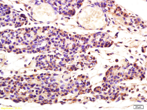
. Aspartate Aminotransferase/GOT1 was detected in paraffin-embedded section of mouse brain tissue. Heat mediated antigen retrieval was performed in EDTA buffer (pH8.0, epitope retrieval solution). The tissue section was blocked with 10% goat serum. The tissue section was then incubated with 2microg/ml rabbit anti-Aspartate Aminotransferase/GOT1 Antibody (A04085-3) overnight at 4°C. Biotinylated goat anti-rabbit IgG was used as secondary antibody and incubated for 30 minutes at 37°C. The tissue section was developed using Strepavidin-Biotin-Complex (SABC) (Catalog # SA1022) with DAB as the chromogen.")
. Aspartate Aminotransferase/GOT1 was detected in paraffin-embedded section of rat brain tissue. Heat mediated antigen retrieval was performed in EDTA buffer (pH8.0, epitope retrieval solution). The tissue section was blocked with 10% goat serum. The tissue section was then incubated with 2microg/ml rabbit anti-Aspartate Aminotransferase/GOT1 Antibody (A04085-3) overnight at 4°C. Biotinylated goat anti-rabbit IgG was used as secondary antibody and incubated for 30 minutes at 37°C. The tissue section was developed using Strepavidin-Biotin-Complex (SABC) (Catalog # SA1022) with DAB as the chromogen.")
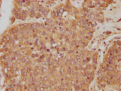
. Aspartate Aminotransferase/GOT1 was detected in immunocytochemical section of T-47D cells. Enzyme antigen retrieval was performed using IHC enzyme antigen retrieval reagent (AR0022) for 15 mins. The cells were blocked with 10% goat serum. And then incubated with 5microg/mL rabbit anti-Aspartate Aminotransferase/GOT1 Antibody (A04085-3) overnight at 4°C. DyLight®488 Conjugated Goat Anti-Rabbit IgG (BA1127) was used as secondary antibody at 1:100 dilution and incubated for 30 minutes at 37°C. The section was counterstained with DAPI. Visualize using a fluorescence microscope and filter sets appropriate for the label used.")
. Overlay histogram showing HepG2 cells stained with A04085-3 (Blue line). To facilitate intracellular staining, cells were fixed with 4% paraformaldehyde and permeabilized with permeabilization buffer. The cells were blocked with 10% normal goat serum. And then incubated with rabbit anti-Aspartate Aminotransferase/GOT1 Antibody (A04085-3, 1microg/1x106 cells) for 30 min at 20°C. DyLight®488 conjugated goat anti-rabbit IgG (BA1127, 5-10microg/1x106 cells) was used as secondary antibody for 30 minutes at 20°C. Isotype control antibody (Green line) was rabbit IgG (1microg/1x106) used under the same conditions. Unlabelled sample without incubation with primary antibody and secondary antibody (Red line) was used as a blank control.")
Figure 1. Western blot analysis of Aspartate Aminotransferase/GOT1 using anti-Aspartate Aminotransferase/GOT1 antibody (A04085-3). Electrophoresis was performed on a 5-20% SDS-PAGE gel at 70V (Stacking gel) / 90V (Resolving gel) for 2-3 hours. The sample well of each lane was loaded with 30ug of sample under reducing conditions. Lane 1: human placenta tissue lysates, Lane 2: human T-47D whole cell lysates, Lane 3: human HepG2 whole cell lysates, Lane 4: human Caco-2 whole cell lysates, Lane 5: human HL-60 whole cell lysates, Lane 6: human K562 whole cell lysates, Lane 7: human Hela whole cell lysates, Lane 8: rat brain tissue lysates, Lane 9: mouse brain tissue lysates, Lane 10: mouse liver tissue lysates. After Electrophoresis, proteins were transferred to a Nitrocellulose membrane at 150mA for 50-90 minutes. Blocked the membrane with 5% Non-fat Milk/ TBS for 1.5 hour at RT. The membrane was incubated with rabbit anti-Aspartate Aminotransferase/GOT1 antigen affinity purified polyclonal antibody (Catalog # A04085-3) at 0.25 microg/mL overnight at 4°C, then washed with TBS-0.1%Tween 3 times with 5 minutes each and probed with a goat anti-rabbit IgG-HRP secondary antibody at a dilution of 1:5000 for 1.5 hour at RT. The signal is developed using an Enhanced Chemiluminescent detection (ECL) kit (Catalog # EK1002) with Tanon 5200 system. A specific band was detected for Aspartate Aminotransferase/GOT1 at approximately 41KD. The expected band size for Aspartate Aminotransferase/GOT1 is at 46KD.
Anti-Aspartate Aminotransferase/GOT1 Antibody Picoband(r)
A04085-3-CARRIER-FREE
ApplicationsFlow Cytometry, ImmunoFluorescence, Western Blot, ELISA, ImmunoCytoChemistry, ImmunoHistoChemistry
Product group Antibodies
ReactivityHuman, Mouse, Rat
TargetGOT1
Overview
- SupplierBoster Bio
- Product NameAnti-Aspartate Aminotransferase/GOT1 Antibody Picoband(r)
- Delivery Days Customer9
- ApplicationsFlow Cytometry, ImmunoFluorescence, Western Blot, ELISA, ImmunoCytoChemistry, ImmunoHistoChemistry
- CertificationResearch Use Only
- ClonalityPolyclonal
- Concentration500 ug/ml
- Gene ID2805
- Target nameGOT1
- Target descriptionglutamic-oxaloacetic transaminase 1
- Target synonymsAST, AST1, ASTQTL1, GIG18, SGOT, cAspAT, cCAT, aspartate aminotransferase, cytoplasmic, aspartate aminotransferase 1, aspartate transaminase 1, cysteine aminotransferase, cytoplasmic, cysteine transaminase, cytoplasmic, glutamate oxaloacetate transaminase 1, glutamic-oxaloacetic transaminase 1, soluble, growth-inhibiting protein 18, testis secretory sperm-binding protein Li 196a, transaminase A
- HostRabbit
- IsotypeIgG
- Protein IDP17174
- Protein NameAspartate aminotransferase, cytoplasmic
- Scientific DescriptionBoster Bio Anti-Aspartate Aminotransferase/GOT1 Antibody Picoband® catalog # A04085-3. Tested in ELISA, Flow Cytometry, IF, IHC, ICC, WB applications. This antibody reacts with Human, Mouse, Rat. The brand Picoband indicates this is a premium antibody that guarantees superior quality, high affinity, and strong signals with minimal background in Western blot applications. Only our best-performing antibodies are designated as Picoband, ensuring unmatched performance.
- ReactivityHuman, Mouse, Rat
- Storage Instruction-20°C,2°C to 8°C
- UNSPSC12352203
Related products
Product group Antibodies
ApplicationsWestern Blot, ELISA
ReactivityHuman
- SizePrice
Product group Antibodies

Goat anti-GOT1 (aa 22-35)EB08132
ApplicationsWestern Blot, ELISA
ReactivityHuman, Mouse
TargetGOT1
- SizePrice
Product group Antibodies
GOT1 AntibodyCSB-PA009679HA01HU
ApplicationsImmunoFluorescence, Western Blot, ELISA, ImmunoHistoChemistry
ReactivityHuman, Mouse
TargetGOT1
- SizePrice

Product group Antibodies
Anti-GOT1 AntibodyHPA064532
ApplicationsWestern Blot, ImmunoHistoChemistry
ReactivityHuman
TargetGOT1
- SizePrice
Product group Antibodies

Aspartate Aminotransferase AntibodyLS-C404720
ApplicationsWestern Blot, ELISA, ImmunoHistoChemistry
ReactivityHuman, Mouse, Rat
TargetGOT1
- SizePrice
Product group Antibodies
GOT1 Polyclonal AntibodyCAC13746
ApplicationsImmunoFluorescence, Western Blot, ELISA, ImmunoHistoChemistry
ReactivityMouse
TargetGOT1
- SizePrice
Product group Antibodies
ApplicationsImmunoFluorescence, Western Blot, ELISA, ImmunoCytoChemistry, ImmunoHistoChemistry, ImmunoHistoChemistry Frozen, ImmunoHistoChemistry Paraffin
ReactivityBovine, Canine, Human, Mouse, Porcine, Rabbit, Rat
TargetGOT1
- SizePrice
![Various tissue extracts (30 μg) were separated by 10% SDS-PAGE, and the membrane was blotted with GOT1 antibody [GT638] (GTX632031) diluted at 1:5000. The HRP-conjugated anti-mouse IgG antibody (GTX213111-01) was used to detect the primary antibody.](https://www.genetex.com/upload/website/prouct_img/normal/GTX632031/GTX632031_41918_20180622_WB_M_tissue_w_23061202_960.webp)
Product group Antibodies
GOT1 antibody [GT638]GTX632031
ApplicationsWestern Blot
ReactivityHuman, Mouse
TargetGOT1
- SizePrice
Product group Antibodies
Anti-GOT1 Antibody144-05822
ApplicationsImmunoFluorescence, Western Blot, ImmunoHistoChemistry
ReactivityHuman, Mouse, Rat
TargetGOT1
- SizePrice