


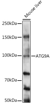
Figure 1. Western blot analysis of ATG9A using anti-ATG9A antibody (A03757-2). Electrophoresis was performed on a 5-20% SDS-PAGE gel at 70V (Stacking gel) / 90V (Resolving gel) for 2-3 hours. The sample well of each lane was loaded with 50ug of sample under reducing conditions. Lane 1: human HepG2 whole cell lysates, Lane 2: human A549 whole cell lysates, Lane 3: human A375 whole cell lysates, Lane 4: human Mcf-7 whole cell lysates, Lane 5: human K562 whole cell lysates, Lane 6: mouse testis tissue lysates. After Electrophoresis, proteins were transferred to a Nitrocellulose membrane at 150mA for 50-90 minutes. Blocked the membrane with 5% Non-fat Milk/ TBS for 1.5 hour at RT. The membrane was incubated with rabbit anti-ATG9A antigen affinity purified polyclonal antibody (Catalog # A03757-2) at 0.5 microg/mL overnight at 4°C, then washed with TBS-0.1%Tween 3 times with 5 minutes each and probed with a goat anti-rabbit IgG-HRP secondary antibody at a dilution of 1:5000 for 1.5 hour at RT. The signal is developed using an Enhanced Chemiluminescent detection (ECL) kit (Catalog # EK1002) with Tanon 5200 system. A specific band was detected for ATG9A at approximately 94KD. The expected band size for ATG9A is at 94KD.
. ATG9A was detected in immunocytochemical section of A549 cells. Enzyme antigen retrieval was performed using IHC enzyme antigen retrieval reagent (AR0022) for 15 mins. The cells were blocked with 10% goat serum. And then incubated with 5microg/mL rabbit anti-ATG9A Antibody (A03757-2) overnight at 4°C. DyLight®488 Conjugated Goat Anti-Rabbit IgG (BA1127) was used as secondary antibody at 1:100 dilution and incubated for 30 minutes at 37°C. The section was counterstained with DAPI. Visualize using a fluorescence microscope and filter sets appropriate for the label used.")
. Overlay histogram showing U87 cells stained with A03757-2 (Blue line). To facilitate intracellular staining, cells were fixed with 4% paraformaldehyde and permeabilized with permeabilization buffer. The cells were blocked with 10% normal goat serum. And then incubated with rabbit anti-ATG9A Antibody (A03757-2, 1microg/1x106 cells) for 30 min at 20°C. DyLight®488 conjugated goat anti-rabbit IgG (BA1127, 5-10microg/1x106 cells) was used as secondary antibody for 30 minutes at 20°C. Isotype control antibody (Green line) was rabbit IgG (1microg/1x106) used under the same conditions. Unlabelled sample without incubation with primary antibody and secondary antibody (Red line) was used as a blank control.")
Figure 1. Western blot analysis of ATG9A using anti-ATG9A antibody (A03757-2). Electrophoresis was performed on a 5-20% SDS-PAGE gel at 70V (Stacking gel) / 90V (Resolving gel) for 2-3 hours. The sample well of each lane was loaded with 50ug of sample under reducing conditions. Lane 1: human HepG2 whole cell lysates, Lane 2: human A549 whole cell lysates, Lane 3: human A375 whole cell lysates, Lane 4: human Mcf-7 whole cell lysates, Lane 5: human K562 whole cell lysates, Lane 6: mouse testis tissue lysates. After Electrophoresis, proteins were transferred to a Nitrocellulose membrane at 150mA for 50-90 minutes. Blocked the membrane with 5% Non-fat Milk/ TBS for 1.5 hour at RT. The membrane was incubated with rabbit anti-ATG9A antigen affinity purified polyclonal antibody (Catalog # A03757-2) at 0.5 microg/mL overnight at 4°C, then washed with TBS-0.1%Tween 3 times with 5 minutes each and probed with a goat anti-rabbit IgG-HRP secondary antibody at a dilution of 1:5000 for 1.5 hour at RT. The signal is developed using an Enhanced Chemiluminescent detection (ECL) kit (Catalog # EK1002) with Tanon 5200 system. A specific band was detected for ATG9A at approximately 94KD. The expected band size for ATG9A is at 94KD.
Anti-ATG9A Antibody Picoband(r)
A03757-2-CARRIER-FREE
ApplicationsFlow Cytometry, ImmunoFluorescence, Western Blot, ELISA, ImmunoCytoChemistry
Product group Antibodies
ReactivityHuman, Mouse
TargetATG9A
Overview
- SupplierBoster Bio
- Product NameAnti-ATG9A Antibody Picoband(r)
- Delivery Days Customer9
- ApplicationsFlow Cytometry, ImmunoFluorescence, Western Blot, ELISA, ImmunoCytoChemistry
- CertificationResearch Use Only
- ClonalityPolyclonal
- Concentration500 ug/ml
- Gene ID79065
- Target nameATG9A
- Target descriptionautophagy related 9A
- Target synonymsAPG9L1, MGD3208, mATG9, autophagy-related protein 9A, APG9 autophagy 9-like 1, APG9-like 1, ATG9 autophagy related 9 homolog A, autophagy 9-like 1 protein
- HostRabbit
- IsotypeIgG
- Protein IDQ7Z3C6
- Protein NameAutophagy-related protein 9A
- Scientific DescriptionBoster Bio Anti-ATG9A Antibody catalog # A03757-2. Tested in ELISA, Flow Cytometry, IF, ICC, WB applications. This antibody reacts with Human, Mouse. The brand Picoband indicates this is a premium antibody that guarantees superior quality, high affinity, and strong signals with minimal background in Western blot applications. Only our best-performing antibodies are designated as Picoband, ensuring unmatched performance.
- ReactivityHuman, Mouse
- Storage Instruction-20°C,2°C to 8°C
- UNSPSC12352203
Related products
Product group Antibodies
Anti-ATG9A AntibodyA16030
ApplicationsWestern Blot
ReactivityHuman, Mouse, Rat
- SizePrice
Product group Antibodies
Anti-ATG9A Antibody144-07994
ApplicationsWestern Blot
ReactivityHuman, Mouse, Rat
TargetATG9A
- SizePrice
Product group Antibodies
ATG9A Recombinant AntibodyBSM-60696R
ApplicationsImmunoFluorescence, Western Blot, ImmunoCytoChemistry, ImmunoHistoChemistry, ImmunoHistoChemistry Frozen, ImmunoHistoChemistry Paraffin
ReactivityHuman
TargetATG9A
- SizePrice
Product group Antibodies
ATG9A AntibodyCSB-PA554014
ApplicationsWestern Blot, ELISA, ImmunoHistoChemistry
ReactivityHuman, Rat
TargetATG9A
- SizePrice
Product group Antibodies

ATG9A AntibodyLS-C401391
ApplicationsELISA, ImmunoHistoChemistry
ReactivityHuman, Rat
TargetATG9A
- SizePrice

Product group Antibodies
Anti-ATG9A AntibodyHPA059551
ApplicationsImmunoCytoChemistry, ImmunoHistoChemistry
ReactivityHuman
TargetATG9A
- SizePrice
![ATG9A antibody detects ATG9A protein at cytoplasm by immunofluorescent analysis. Sample: HeLa cells were fixed in ice-cold MeOH for 5 min. Green: ATG9A stained by ATG9A antibody (GTX128427) diluted at 1:500. Red: alpha Tubulin, a cytoskeleton marker, stained by alpha Tubulin antibody [GT114] (GTX628802) diluted at 1:1000. Blue: Fluoroshield with DAPI (GTX30920).](https://www.genetex.com/upload/website/prouct_img/normal/GTX128427/GTX128427_44545_20220429_ICC_IF_w_23060523_217.webp)
Product group Antibodies
ATG9A antibodyGTX128427
ApplicationsImmunoFluorescence, ImmunoPrecipitation, Western Blot, ImmunoCytoChemistry, ImmunoHistoChemistry, ImmunoHistoChemistry Paraffin
ReactivityHuman, Mouse, Rat
TargetATG9A
- SizePrice