


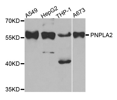
Figure 1. Western blot analysis of ATGL/PNPLA2 using anti-ATGL/PNPLA2 antibody (M01800). Electrophoresis was performed on a 5-20% SDS-PAGE gel at 70V (Stacking gel) / 90V (Resolving gel) for 2-3 hours. The sample well of each lane was loaded with 30 ug of sample under reducing conditions. Lane 1: human A431 whole cell lysates, Lane 2: human 293T whole cell lysates, Lane 3: human SiHa whole cell lysates, Lane 4: human HepG2 whole cell lysates, Lane 5: rat C6 whole cell lysates, Lane 6: rat heart tissue lysates, Lane 7: mouse NIH/3T3 whole cell lysates, Lane 8: mouse heart tissue lysates. After electrophoresis, proteins were transferred to a nitrocellulose membrane at 150 mA for 50-90 minutes. Blocked the membrane with 5% non-fat milk/TBS for 1.5 hour at RT. The membrane was incubated with rabbit anti-ATGL/PNPLA2 antigen affinity purified monoclonal antibody (Catalog # M01800) at 1:500 overnight at 4°C, then washed with TBS-0.1%Tween 3 times with 5 minutes each and probed with a goat anti-rabbit IgG-HRP secondary antibody at a dilution of 1:1000 for 1.5 hour at RT. The signal is developed using an Enhanced Chemiluminescent detection (ECL) kit (Catalog # EK1002) with Tanon 5200 system. A specific band was detected for ATGL/PNPLA2 at approximately 55 kDa. The expected band size for ATGL/PNPLA2 is at 55 kDa.
Figure 1. Western blot analysis of ATGL/PNPLA2 using anti-ATGL/PNPLA2 antibody (M01800). Electrophoresis was performed on a 5-20% SDS-PAGE gel at 70V (Stacking gel) / 90V (Resolving gel) for 2-3 hours. The sample well of each lane was loaded with 30 ug of sample under reducing conditions. Lane 1: human A431 whole cell lysates, Lane 2: human 293T whole cell lysates, Lane 3: human SiHa whole cell lysates, Lane 4: human HepG2 whole cell lysates, Lane 5: rat C6 whole cell lysates, Lane 6: rat heart tissue lysates, Lane 7: mouse NIH/3T3 whole cell lysates, Lane 8: mouse heart tissue lysates. After electrophoresis, proteins were transferred to a nitrocellulose membrane at 150 mA for 50-90 minutes. Blocked the membrane with 5% non-fat milk/TBS for 1.5 hour at RT. The membrane was incubated with rabbit anti-ATGL/PNPLA2 antigen affinity purified monoclonal antibody (Catalog # M01800) at 1:500 overnight at 4°C, then washed with TBS-0.1%Tween 3 times with 5 minutes each and probed with a goat anti-rabbit IgG-HRP secondary antibody at a dilution of 1:1000 for 1.5 hour at RT. The signal is developed using an Enhanced Chemiluminescent detection (ECL) kit (Catalog # EK1002) with Tanon 5200 system. A specific band was detected for ATGL/PNPLA2 at approximately 55 kDa. The expected band size for ATGL/PNPLA2 is at 55 kDa.
Anti-ATGL / PNPLA2 Rabbit Monoclonal Antibody
M01800
ApplicationsWestern Blot
Product group Antibodies
ReactivityHuman, Mouse, Rat
TargetPNPLA2
Overview
- SupplierBoster Bio
- Product NameAnti-ATGL / PNPLA2 Rabbit Monoclonal Antibody
- Delivery Days Customer9
- ApplicationsWestern Blot
- CertificationResearch Use Only
- ClonalityMonoclonal
- Clone ID23P38
- Gene ID57104
- Target namePNPLA2
- Target descriptionpatatin like domain 2, triacylglycerol lipase
- Target synonyms1110001C14Rik, ATGL, FP17548, PEDF-R, TTS-2.2, TTS2, iPLA2zeta, patatin-like phospholipase domain-containing protein 2, IPLA2-zeta, TTS2.2, adipose triglyceride lipase, calcium-independent phospholipase A2, calcium-independent phospholipase A2-zeta, desnutrin, mutant patatin-like phospholipase domain containing 2, patatin like phospholipase domain containing 2, pigment epithelium-derived factor, pigment epithelium-derived factor receptor, transport-secretion protein 2.2, triglyceride hydrolase
- HostRabbit
- IsotypeIgG
- Protein IDQ96AD5
- Protein NamePatatin-like phospholipase domain-containing protein 2
- Scientific DescriptionBoster Bio Anti-ATGL / PNPLA2 Rabbit Monoclonal Antibody catalog # M01800. Tested in WB application. This antibody reacts with Human, Mouse, Rat.
- ReactivityHuman, Mouse, Rat
- Storage Instruction-20°C
- UNSPSC12352203
Related products
Product group Antibodies
Anti-PNPLA2 AntibodyA31175
ApplicationsWestern Blot, ImmunoHistoChemistry
ReactivityHuman, Mouse, Rat
- SizePrice

Product group Antibodies
ApplicationsFlow Cytometry, ImmunoFluorescence, Western Blot, ELISA, ImmunoCytoChemistry, ImmunoHistoChemistry, ImmunoHistoChemistry Frozen, ImmunoHistoChemistry Paraffin
ReactivityHuman, Mouse, Rat
TargetPNPLA2
- SizePrice
Product group Antibodies

Goat anti-PNPLA2 / ATGLEB07640
ApplicationsWestern Blot, ELISA
ReactivityHuman
TargetPNPLA2
- SizePrice
Product group Antibodies
PNPLA2 AntibodyCSB-PA836180ESR1HU
ApplicationsWestern Blot, ELISA, ImmunoHistoChemistry
ReactivityHuman
TargetPNPLA2
- SizePrice
Product group Antibodies
Pnpla2 Polyclonal AntibodyCAC07568
ApplicationsImmunoFluorescence, Western Blot, ELISA, ImmunoHistoChemistry
ReactivityMouse
TargetPNPLA2
- SizePrice
Product group Antibodies

PNPLA2 / ATGL AntibodyLS-C334584
ApplicationsWestern Blot, ImmunoHistoChemistry
ReactivityHuman, Mouse, Rat
TargetPNPLA2
- SizePrice

Product group Antibodies
Anti-PNPLA2 AntibodyHPA055173
ApplicationsImmunoCytoChemistry
ReactivityHuman
TargetPNPLA2
- SizePrice
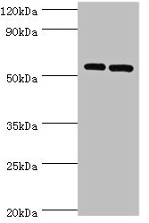
![Mouse tissue extract (50 μg) was separated by 7.5% SDS-PAGE, and the membrane was blotted with ATGL antibody [N1C1] (GTX109941) diluted at 1:1000. The HRP-conjugated anti-rabbit IgG antibody (GTX213110-01) was used to detect the primary antibody.](https://www.genetex.com/upload/website/prouct_img/normal/GTX109941/GTX109941_42466_20170420_WB_M_white_adipose_w_23060500_208.webp)
Product group Antibodies
ATGL antibody [N1C1]GTX109941
ApplicationsWestern Blot
ReactivityHuman, Mouse
TargetPNPLA2
- SizePrice
Product group Antibodies
TargetPNPLA2
- SizePrice
Product group Antibodies
Anti-PNPLA2Y158256
ApplicationsWestern Blot, ELISA, ImmunoHistoChemistry
ReactivityHuman
- SizePrice