


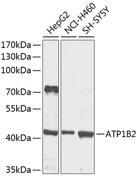
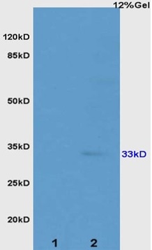
Figure 1. Western blot analysis of ATP1B2 using anti-ATP1B2 antibody (A07027-2). Electrophoresis was performed on a 5-20% SDS-PAGE gel at 70V (Stacking gel) / 90V (Resolving gel) for 2-3 hours. The sample well of each lane was loaded with 30 ug of sample under reducing conditions. Lane 1: rat brain tissue lysates, Lane 2: mouse brain tissue lysates. After electrophoresis, proteins were transferred to a nitrocellulose membrane at 150 mA for 50-90 minutes. Blocked the membrane with 5% non-fat milk/TBS for 1.5 hour at RT. The membrane was incubated with rabbit anti-ATP1B2 antigen affinity purified polyclonal antibody (Catalog # A07027-2) at 0.5 microg/mL overnight at 4°C, then washed with TBS-0.1%Tween 3 times with 5 minutes each and probed with a goat anti-rabbit IgG-HRP secondary antibody at a dilution of 1:5000 for 1.5 hour at RT. The signal is developed using an Enhanced Chemiluminescent detection (ECL) kit (Catalog # EK1002) with Tanon 5200 system. A specific band was detected for ATP1B2 at approximately 44 kDa. The expected band size for ATP1B2 is at 33 kDa.
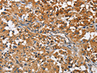
. ATP1B2 was detected in a paraffin-embedded section of human glioma tissue. Heat mediated antigen retrieval was performed in EDTA buffer (pH 8.0, epitope retrieval solution). The tissue section was blocked with 10% goat serum. The tissue section was then incubated with 2 microg/ml rabbit anti-ATP1B2 Antibody (A07027-2) overnight at 4°C. Peroxidase Conjugated Goat Anti-rabbit IgG was used as secondary antibody and incubated for 30 minutes at 37°C. The tissue section was developed using HRP Conjugated Rabbit IgG Super Vision Assay Kit (Catalog # SV0002) with DAB as the chromogen.")
. Overlay histogram showing U87 cells stained with A07027-2 (Blue line). The cells were fixed with 4% paraformaldehyde and blocked with 10% normal goat serum. And then incubated with rabbit anti-ATP1B2 Antibody (A07027-2, 1 microg/1x106 cells) for 30 min at 20°C. DyLight®488 conjugated goat anti-rabbit IgG (BA1127, 5-10 microg/1x106 cells) was used as secondary antibody for 30 minutes at 20°C. Isotype control antibody (Green line) was rabbit IgG (1 microg/1x106) used under the same conditions. Unlabelled sample without incubation with primary antibody and secondary antibody (Red line) was used as a blank control.")
Figure 1. Western blot analysis of ATP1B2 using anti-ATP1B2 antibody (A07027-2). Electrophoresis was performed on a 5-20% SDS-PAGE gel at 70V (Stacking gel) / 90V (Resolving gel) for 2-3 hours. The sample well of each lane was loaded with 30 ug of sample under reducing conditions. Lane 1: rat brain tissue lysates, Lane 2: mouse brain tissue lysates. After electrophoresis, proteins were transferred to a nitrocellulose membrane at 150 mA for 50-90 minutes. Blocked the membrane with 5% non-fat milk/TBS for 1.5 hour at RT. The membrane was incubated with rabbit anti-ATP1B2 antigen affinity purified polyclonal antibody (Catalog # A07027-2) at 0.5 microg/mL overnight at 4°C, then washed with TBS-0.1%Tween 3 times with 5 minutes each and probed with a goat anti-rabbit IgG-HRP secondary antibody at a dilution of 1:5000 for 1.5 hour at RT. The signal is developed using an Enhanced Chemiluminescent detection (ECL) kit (Catalog # EK1002) with Tanon 5200 system. A specific band was detected for ATP1B2 at approximately 44 kDa. The expected band size for ATP1B2 is at 33 kDa.
Anti-ATP1B2 Antibody Picoband(r)
A07027-2-CARRIER-FREE
ApplicationsFlow Cytometry, Western Blot, ELISA, ImmunoHistoChemistry
Product group Antibodies
ReactivityHuman, Mouse, Rat
TargetATP1B2
Overview
- SupplierBoster Bio
- Product NameAnti-ATP1B2 Antibody Picoband(r)
- Delivery Days Customer9
- ApplicationsFlow Cytometry, Western Blot, ELISA, ImmunoHistoChemistry
- CertificationResearch Use Only
- ClonalityPolyclonal
- Concentration500 ug/ml
- Gene ID482
- Target nameATP1B2
- Target descriptionATPase Na+/K+ transporting subunit beta 2
- Target synonymsAMOG, sodium/potassium-transporting ATPase subunit beta-2, ATPase, Na+/K+ transporting, beta 2 polypeptide, Na, K-ATPase beta-2 polypeptide, adhesion molecule in glia, adhesion molecule on glia, sodium pump subunit beta-2, sodium-potassium ATPase subunit beta 2 (non-catalytic), sodium/potassium-dependent ATPase beta-2 subunit, sodium/potassium-dependent ATPase subunit beta-2, sodium/potassium-transporting ATPase beta-2 chain
- HostRabbit
- IsotypeIgG
- Protein IDP14415
- Protein NameSodium/potassium-transporting ATPase subunit beta-2
- Scientific DescriptionBoster Bio Anti-ATP1B2 Antibody Picoband® catalog # A07027-2. Tested in ELISA, Flow Cytometry, IHC, WB applications. This antibody reacts with Human, Mouse, Rat. The brand Picoband indicates this is a premium antibody that guarantees superior quality, high affinity, and strong signals with minimal background in Western blot applications. Only our best-performing antibodies are designated as Picoband, ensuring unmatched performance.
- ReactivityHuman, Mouse, Rat
- Storage Instruction-20°C,2°C to 8°C
- UNSPSC12352203
Related products
Product group Antibodies
ATP1B2 AntibodyCSB-PA586126
ApplicationsELISA, ImmunoHistoChemistry
ReactivityHuman, Mouse, Rat
TargetATP1B2
- SizePrice
Product group Antibodies
Anti-ATP1B2 AntibodyA16373
ApplicationsImmunoFluorescence, Western Blot, ImmunoCytoChemistry
ReactivityHuman, Mouse, Rat
- SizePrice
Product group Antibodies

AMOG / ATP1B2 AntibodyLS-C831142
ApplicationsWestern Blot, ELISA, ImmunoHistoChemistry
ReactivityHuman, Mouse, Rat
TargetATP1B2
- SizePrice

Product group Antibodies
Anti-ATP1B2 AntibodyHPA010698
ApplicationsImmunoHistoChemistry
ReactivityHuman
TargetATP1B2
- SizePrice
Product group Antibodies
Anti-ATP1B2 Antibody144-09928
ApplicationsWestern Blot
ReactivityHuman, Mouse, Rat
TargetATP1B2
- SizePrice
Product group Antibodies
References
ATP1B2 Polyclonal AntibodyBS-1152R
ApplicationsImmunoFluorescence, Western Blot, ELISA, ImmunoCytoChemistry, ImmunoHistoChemistry, ImmunoHistoChemistry Frozen, ImmunoHistoChemistry Paraffin
ReactivityHuman, Mouse, Rat
TargetATP1B2
- SizePrice