


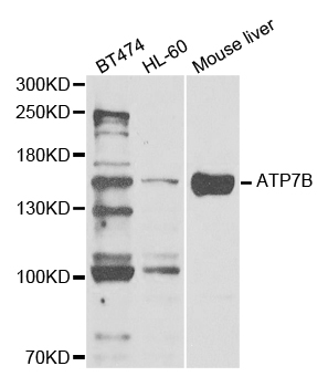
Figure 1. Western blot analysis of ATP7B using anti-ATP7B antibody (A00686-2). Electrophoresis was performed on a 5-20% SDS-PAGE gel at 70V (Stacking gel) / 90V (Resolving gel) for 2-3 hours. The sample well of each lane was loaded with 30 ug of sample under reducing conditions. Lane 1: human HepG2 whole cell lysates, Lane 2: rat liver tissue lysates, Lane 3: mouse kidney tissue lysates. After electrophoresis, proteins were transferred to a nitrocellulose membrane at 150 mA for 50-90 minutes. Blocked the membrane with 5% non-fat milk/TBS for 1.5 hour at RT. The membrane was incubated with rabbit anti-ATP7B antigen affinity purified polyclonal antibody (Catalog # A00686-2) at 0.5 microg/mL overnight at 4°C, then washed with TBS-0.1%Tween 3 times with 5 minutes each and probed with a goat anti-rabbit IgG-HRP secondary antibody at a dilution of 1:5000 for 1.5 hour at RT. The signal is developed using an Enhanced Chemiluminescent detection (ECL) kit (Catalog # EK1002) with Tanon 5200 system. A specific band was detected for ATP7B at approximately 157 kDa. The expected band size for ATP7B is at 157 kDa.
. ATP7B was detected in an immunocytochemical section of Caco-2 cells. Enzyme antigen retrieval was performed using IHC enzyme antigen retrieval reagent (AR0022) for 15 mins. The cells were blocked with 10% goat serum. And then incubated with 5 microg/mL rabbit anti-ATP7B Antibody (A00686-2) overnight at 4°C. DyLight®488 Conjugated Goat Anti-Rabbit IgG (BA1127) was used as secondary antibody at 1:100 dilution and incubated for 30 minutes at 37°C. The section was counterstained with DAPI. Visualize using a fluorescence microscope and filter sets appropriate for the label used.")
Figure 1. Western blot analysis of ATP7B using anti-ATP7B antibody (A00686-2). Electrophoresis was performed on a 5-20% SDS-PAGE gel at 70V (Stacking gel) / 90V (Resolving gel) for 2-3 hours. The sample well of each lane was loaded with 30 ug of sample under reducing conditions. Lane 1: human HepG2 whole cell lysates, Lane 2: rat liver tissue lysates, Lane 3: mouse kidney tissue lysates. After electrophoresis, proteins were transferred to a nitrocellulose membrane at 150 mA for 50-90 minutes. Blocked the membrane with 5% non-fat milk/TBS for 1.5 hour at RT. The membrane was incubated with rabbit anti-ATP7B antigen affinity purified polyclonal antibody (Catalog # A00686-2) at 0.5 microg/mL overnight at 4°C, then washed with TBS-0.1%Tween 3 times with 5 minutes each and probed with a goat anti-rabbit IgG-HRP secondary antibody at a dilution of 1:5000 for 1.5 hour at RT. The signal is developed using an Enhanced Chemiluminescent detection (ECL) kit (Catalog # EK1002) with Tanon 5200 system. A specific band was detected for ATP7B at approximately 157 kDa. The expected band size for ATP7B is at 157 kDa.
Anti-ATP7B Antibody Picoband(r)
A00686-2-CARRIER-FREE
ApplicationsImmunoFluorescence, Western Blot, ELISA, ImmunoCytoChemistry
Product group Antibodies
ReactivityHuman, Mouse, Rat
TargetATP7B
Overview
- SupplierBoster Bio
- Product NameAnti-ATP7B Antibody Picoband(r)
- Delivery Days Customer9
- ApplicationsImmunoFluorescence, Western Blot, ELISA, ImmunoCytoChemistry
- CertificationResearch Use Only
- ClonalityPolyclonal
- Concentration500 ug/ml
- Gene ID540
- Target nameATP7B
- Target descriptionATPase copper transporting beta
- Target synonymsPWD, WC1, WD, WND, copper-transporting ATPase 2, ATPase, Cu(2+)- transporting, beta polypeptide, ATPase, Cu++ transporting, beta polypeptide, Wilson disease-associated protein, copper pump 2, copper-transporting protein ATP7B
- HostRabbit
- IsotypeIgG
- Protein IDP35670
- Protein NameCopper-transporting ATPase 2
- Scientific DescriptionBoster Bio Anti-ATP7B Antibody Picoband® catalog # A00686-2. Tested in ELISA, IF, ICC, WB applications. This antibody reacts with Human, Mouse, Rat. The brand Picoband indicates this is a premium antibody that guarantees superior quality, high affinity, and strong signals with minimal background in Western blot applications. Only our best-performing antibodies are designated as Picoband, ensuring unmatched performance.
- ReactivityHuman, Mouse, Rat
- Storage Instruction-20°C,2°C to 8°C
- UNSPSC12352203
Related products
Product group Antibodies
Anti-ATP7B AntibodyA30858
ApplicationsImmunoFluorescence, Western Blot, ImmunoHistoChemistry
ReactivityHuman, Mouse, Rat
- SizePrice
Product group Antibodies
Anti-ATP7B Antibody144-05676
ApplicationsImmunoFluorescence, Western Blot
ReactivityHuman, Mouse
TargetATP7B
- SizePrice
Product group Antibodies
ATP7B AntibodyCSB-PA002415ESR2HU
ApplicationsELISA, ImmunoHistoChemistry
ReactivityHuman
TargetATP7B
- SizePrice
Product group Antibodies
Atp7B Polyclonal AntibodyCAC10561
ApplicationsELISA, ImmunoHistoChemistry
TargetATP7B
- SizePrice
Product group Antibodies
References
ATP7B Polyclonal AntibodyBS-1718R
ApplicationsImmunoFluorescence, Western Blot, ELISA, ImmunoCytoChemistry, ImmunoHistoChemistry, ImmunoHistoChemistry Frozen, ImmunoHistoChemistry Paraffin
ReactivityChicken, Human, Mouse, Porcine, Rabbit, Rat
TargetATP7B
- SizePrice
![ICC/IF analysis of 4%PFA-fixed NIH-3T3 using GTX03698 ATP7b antibody [S62-29]. Green : Primary antibody Blue : DAPI Red : F-Actin Dilution : 1:100](https://www.genetex.com/upload/website/prouct_img/normal/GTX03698/GTX03698_20220518_ICCIF_1_w_23053123_518.webp)
Product group Antibodies
ATP7b antibody [S62-29]GTX03698
ApplicationsImmunoFluorescence, ImmunoPrecipitation, Western Blot, ImmunoCytoChemistry, ImmunoHistoChemistry
ReactivityHuman, Mouse, Rat
TargetATP7B
- SizePrice
Product group Antibodies

WC1 / ATP7B Antibody (N-Terminus)LS-C358914
ApplicationsImmunoFluorescence, Western Blot, ImmunoCytoChemistry, ImmunoHistoChemistry, ImmunoHistoChemistry Paraffin
ReactivityHuman
TargetATP7B
- SizePrice

Product group Antibodies
Anti-ATP7B AntibodyHPA009137
ApplicationsImmunoCytoChemistry
ReactivityHuman
TargetATP7B
- SizePrice