

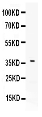
Figure 1. Western blot analysis of B7 DC using anti-B7 DC antibody (PB10010). Electrophoresis was performed on a 5-20% SDS-PAGE gel at 70V (Stacking gel) / 90V (Resolving gel) for 2-3 hours. The sample well of each lane was loaded with 30 ug of sample under reducing conditions. Lane 1: HELA whole cell lysates. After electrophoresis, proteins were transferred to a nitrocellulose membrane at 150 mA for 50-90 minutes. Blocked the membrane with 5% non-fat milk/TBS for 1.5 hour at RT. The membrane was incubated with rabbit anti-B7 DC antigen affinity purified polyclonal antibody (Catalog # PB10010) at 0.5 microg/mL overnight at 4°C, then washed with TBS-0.1%Tween 3 times with 5 minutes each and probed with a goat anti-rabbit IgG-HRP secondary antibody at a dilution of 1:5000 for 1.5 hour at RT. The signal is developed using an Enhanced Chemiluminescent detection (ECL) kit (Catalog # EK1002) with Tanon 5200 system. A specific band was detected for B7 DC at approximately 37 kDa. The expected band size for B7 DC is at 31 kDa.
Figure 1. Western blot analysis of B7 DC using anti-B7 DC antibody (PB10010). Electrophoresis was performed on a 5-20% SDS-PAGE gel at 70V (Stacking gel) / 90V (Resolving gel) for 2-3 hours. The sample well of each lane was loaded with 30 ug of sample under reducing conditions. Lane 1: HELA whole cell lysates. After electrophoresis, proteins were transferred to a nitrocellulose membrane at 150 mA for 50-90 minutes. Blocked the membrane with 5% non-fat milk/TBS for 1.5 hour at RT. The membrane was incubated with rabbit anti-B7 DC antigen affinity purified polyclonal antibody (Catalog # PB10010) at 0.5 microg/mL overnight at 4°C, then washed with TBS-0.1%Tween 3 times with 5 minutes each and probed with a goat anti-rabbit IgG-HRP secondary antibody at a dilution of 1:5000 for 1.5 hour at RT. The signal is developed using an Enhanced Chemiluminescent detection (ECL) kit (Catalog # EK1002) with Tanon 5200 system. A specific band was detected for B7 DC at approximately 37 kDa. The expected band size for B7 DC is at 31 kDa.
Anti-B7 DC/PDCD1LG2 Antibody Picoband(r)
PB10010-CARRIER-FREE
ApplicationsWestern Blot
Product group Antibodies
ReactivityHuman
TargetPDCD1LG2
Overview
- SupplierBoster Bio
- Product NameAnti-B7 DC/PDCD1LG2 Antibody Picoband(r)
- Delivery Days Customer9
- Application Supplier NoteTested Species: In-house tested species with positive results. Other applications have not been tested. Optimal dilutions should be determined by end users.
- ApplicationsWestern Blot
- CertificationResearch Use Only
- ClonalityPolyclonal
- Concentration500 ug/ml
- Gene ID80380
- Target namePDCD1LG2
- Target descriptionprogrammed cell death 1 ligand 2
- Target synonymsB7DC, Btdc, CD273, PD-L2, PDCD1L2, PDL2, bA574F11.2, programmed cell death 1 ligand 2, B7 dendritic cell molecule, B7-DC, PD-1-ligand 2, PDCD1 ligand 2, butyrophilin B7-DC, programmed death ligand 2
- HostRabbit
- IsotypeIgG
- Protein IDQ9BQ51
- Protein NameProgrammed cell death 1 ligand 2
- Scientific DescriptionBoster Bio Anti-B7 DC/PDCD1LG2 Antibody Picoband® catalog # PB10010. Tested in WB applications. This antibody reacts with Human. The brand Picoband indicates this is a premium antibody that guarantees superior quality, high affinity, and strong signals with minimal background in Western blot applications. Only our best-performing antibodies are designated as Picoband, ensuring unmatched performance.
- ReactivityHuman
- Storage Instruction-20°C,2°C to 8°C
- UNSPSC12352203
Related products
Product group Antibodies
Anti-PDCD1LG2 AntibodyA44990
ApplicationsImmunoHistoChemistry
ReactivityHuman
- SizePrice
Product group Antibodies
Anti-PDCD1LG2 (N-term) Antibody102-20342
ApplicationsWestern Blot, ImmunoHistoChemistry, ImmunoHistoChemistry Paraffin
TargetPDCD1LG2
- SizePrice
Product group Antibodies

Anti-PDL2 [Z64P2D3H4]Ab00852-1.1
ApplicationsWestern Blot, ELISA, ImmunoHistoChemistry
ReactivityHuman
TargetPDCD1LG2
- SizePrice
Product group Antibodies
PDCD1LG2 Polyclonal AntibodyBS-1868R
ApplicationsELISA, ImmunoHistoChemistry, ImmunoHistoChemistry Paraffin
ReactivityBovine, Human, Mouse, Rat
TargetPDCD1LG2
- SizePrice
Product group Antibodies
ApplicationsImmunoPrecipitation, Western Blot, ImmunoCytoChemistry, ImmunoHistoChemistry
TargetPDCD1LG2
- SizePrice
Product group Antibodies
PDCD1LG2 AntibodyCSB-PA017667EA01HU
ApplicationsELISA
ReactivityHuman
TargetPDCD1LG2
- SizePrice
Product group Antibodies

PD-L2 / PDCD1LG2 / CD273 AntibodyLS-C404728
ApplicationsELISA, ImmunoHistoChemistry
ReactivityHuman
TargetPDCD1LG2
- SizePrice
Product group Antibodies
References
PD-L2 antibodyGTX85449
ApplicationsWestern Blot, ELISA, ImmunoHistoChemistry, ImmunoHistoChemistry Paraffin
ReactivityHuman, Mouse, Rat
TargetPDCD1LG2
- SizePrice

Product group Antibodies
Anti-PDCD1LG2 AntibodyHPA013411
ApplicationsImmunoCytoChemistry, ImmunoHistoChemistry
ReactivityHuman
TargetPDCD1LG2
- SizePrice