

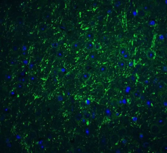
IHC-P analysis of mouse brain tissue using GTX85449 PD-L2 antibody. Working concentration : 20 μg/ml


IHC-P analysis of mouse brain tissue using GTX85449 PD-L2 antibody. Working concentration : 20 μg/ml
PD-L2 antibody
GTX85449
ApplicationsWestern Blot, ELISA, ImmunoHistoChemistry, ImmunoHistoChemistry Paraffin
Product group Antibodies
ReactivityHuman, Mouse, Rat
TargetPDCD1LG2
Overview
- SupplierGeneTex
- Product NamePD-L2 antibody
- Delivery Days Customer9
- Application Supplier NoteWB: 0.5 - 1 microg/mL. IHC-P: 2.5 microg/mL. *Optimal dilutions/concentrations should be determined by the researcher.Not tested in other applications.
- ApplicationsWestern Blot, ELISA, ImmunoHistoChemistry, ImmunoHistoChemistry Paraffin
- CertificationResearch Use Only
- ClonalityPolyclonal
- Concentration1 mg/ml
- ConjugateUnconjugated
- Gene ID80380
- Target namePDCD1LG2
- Target descriptionprogrammed cell death 1 ligand 2
- Target synonymsB7DC, Btdc, CD273, PD-L2, PDCD1L2, PDL2, bA574F11.2, programmed cell death 1 ligand 2, B7 dendritic cell molecule, B7-DC, PD-1-ligand 2, PDCD1 ligand 2, butyrophilin B7-DC, programmed death ligand 2
- HostRabbit
- IsotypeIgG
- Protein IDQ9BQ51
- Protein NameProgrammed cell death 1 ligand 2
- Scientific DescriptionCell-mediated immune responses are initiated by T lymphocytes that are themselves stimulated by co gnate peptides bound to MHC molecules on antigen-presenting cells (APC). T-cell activation is generally self-limited as activated T cells express receptors such as PD-1 (also known as PDCD-1) that mediate inhibitory signals from the APC. PD-1 can bind two different but related ligands, PDL-1 and PDL-2, both of which are thought act as a negative regulator of T cell activation. However, it has been suggested that PDL-2 can act to stimulate an immunogenic response through and alternative receptor from PD-1. At least three isoforms of PDL-2 are known to exist; this antibody is specific to the largest isoform. PDL-2 antibody has no cross-reactivity to PDL-2.
- ReactivityHuman, Mouse, Rat
- Storage Instruction-20°C or -80°C,2°C to 8°C
- UNSPSC41116161
References
- Splenic CD4(+) and CD8(+) T-cells highly expressed PD-1 and Tim-3 in cirrhotic patients with HCV infection and portal hypertension. Huang N et al., 2021 Jan-Dec, Int J Immunopathol PharmacolRead this paper
Datasheet
Related products
Product group Antibodies
Anti-PDCD1LG2 AntibodyA44990
ApplicationsImmunoHistoChemistry
ReactivityHuman
- SizePrice
Product group Antibodies
Anti-PDCD1LG2 (N-term) Antibody102-20342
ApplicationsWestern Blot, ImmunoHistoChemistry, ImmunoHistoChemistry Paraffin
TargetPDCD1LG2
- SizePrice
Product group Antibodies

Anti-PDL2 [Z64P2D3H4]Ab00852-1.1
ApplicationsWestern Blot, ELISA, ImmunoHistoChemistry
ReactivityHuman
TargetPDCD1LG2
- SizePrice
Product group Antibodies
PDCD1LG2 Polyclonal AntibodyBS-1868R
ApplicationsELISA, ImmunoHistoChemistry, ImmunoHistoChemistry Paraffin
ReactivityBovine, Human, Mouse, Rat
TargetPDCD1LG2
- SizePrice
Product group Antibodies
ApplicationsImmunoPrecipitation, Western Blot, ImmunoCytoChemistry, ImmunoHistoChemistry
TargetPDCD1LG2
- SizePrice
Product group Antibodies
PDCD1LG2 AntibodyCSB-PA017667EA01HU
ApplicationsELISA
ReactivityHuman
TargetPDCD1LG2
- SizePrice
Product group Antibodies

PD-L2 / PDCD1LG2 / CD273 AntibodyLS-C404728
ApplicationsELISA, ImmunoHistoChemistry
ReactivityHuman
TargetPDCD1LG2
- SizePrice
Product group Antibodies
PD-L2 antibody [10B6]GTX52964
ApplicationsFlow Cytometry, Western Blot, ImmunoHistoChemistry, ImmunoHistoChemistry Paraffin
ReactivityHuman
TargetPDCD1LG2
- SizePrice

Product group Antibodies
Anti-PDCD1LG2 AntibodyHPA013411
ApplicationsImmunoCytoChemistry, ImmunoHistoChemistry
ReactivityHuman
TargetPDCD1LG2
- SizePrice
Product group Antibodies
Anti-B7 DC/PDCD1LG2 Antibody Picoband(r)PB10010-CARRIER-FREE
ApplicationsWestern Blot
ReactivityHuman
TargetPDCD1LG2
- SizePrice