

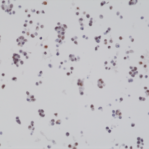
Immunohistochemical staining of formalin fixed and paraffin embedded 22RV1 cell section using anti-BAG-1L rabbit monoclonal antibody (Clone RM310) at a 1:2000 dilution.
Immunohistochemical staining of formalin fixed and paraffin embedded 22RV1 cell section using anti-BAG-1L rabbit monoclonal antibody (Clone RM310) at a 1:2000 dilution.
anti-BAG-1L (human), Rabbit Monoclonal (RM310)
REV-31-1196-00
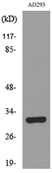
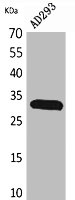
ApplicationsWestern Blot, ImmunoCytoChemistry, ImmunoHistoChemistry
Product group Antibodies
ReactivityHuman
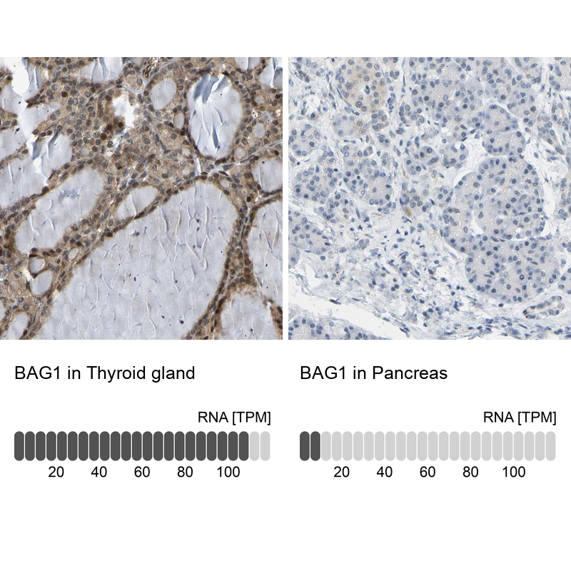
TargetBAG1
Overview
- SupplierRevMAb Biosciences
- Product Nameanti-BAG-1L (human), Rabbit Monoclonal (RM310)
- Delivery Days Customer2
- ApplicationsWestern Blot, ImmunoCytoChemistry, ImmunoHistoChemistry
- CertificationResearch Use Only
- ClonalityMonoclonal
- Clone IDRM310
- Gene ID573
- Target nameBAG1
- Target descriptionBAG cochaperone 1
- Target synonymsBAG-1, HAP, RAP46, BAG family molecular chaperone regulator 1, BCL2 associated athanogene 1, BCL2-associated athanogene, Bcl-2 associating athanogene-1 protein, Bcl-2-binding protein, glucocortoid receptor-associated protein RAP46, receptor-associated protein, 46-KD
- HostRabbit
- IsotypeIgG
- Protein IDQ99933
- Protein NameBAG family molecular chaperone regulator 1
- Scientific DescriptionBAG-1L belongs to a family of evolutionary conserved proteins, containing an approximately 110 amino acid sequence termed the BAG domain that defines the protein family. BAG1 was identified in a screen of interacting proteins of the anti-apoptotic protein BCL2. BAG1 shares no significant homology with BCL2 or other BCL2 family proteins, which can form homo- and heterodimers. However, coexpression of BAG1 and BCL2 provided markedly increased protection from cell death induced by several stimuli. BAG1 itself is made up of different isoforms translated from a single mRNA by alternative translational initiation, resulting in similar carboxy-terminal sequences but different N-terminal sequences. In humans, there are four BAG1 isoforms (BAG1L, 1M, 1 and 1S). The multiple protein isoforms interact with diverse partners, including chaperones Hsc70/Hsp70, Ser/Thr kinase Raf-1 and Bcl-2, to promote cancer cell survival. The BAG-1L isoform specifically binds to and increases the transcriptional activity of oestrogen receptor in cells, and in some, but not all studies, BAG-1 expression is predictive of clinical outcome in breast cancer. - Recombinant Antibody. This antibody reacts to human BAG-1L. It does not cross react to BAG-1M or BAG-1S. Applications: WB, ICC, IHC. Source: Rabbit. Liquid. 50% Glycerol/PBS with 1% BSA and 0.09% sodium azide. BAG-1L belongs to a family of evolutionary conserved proteins, containing an approximately 110 amino acid sequence termed the BAG domain that defines the protein family. BAG1 was identified in a screen of interacting proteins of the anti-apoptotic protein BCL2. BAG1 shares no significant homology with BCL2 or other BCL2 family proteins, which can form homo- and heterodimers. However, coexpression of BAG1 and BCL2 provided markedly increased protection from cell death induced by several stimuli. BAG1 itself is made up of different isoforms translated from a single mRNA by alternative translational initiation, resulting in similar carboxy-terminal sequences but different N-terminal sequences. In humans, there are four BAG1 isoforms (BAG1L, 1M, 1 and 1S). The multiple protein isoforms interact with diverse partners, including chaperones Hsc70/Hsp70, Ser/Thr kinase Raf-1 and Bcl-2, to promote cancer cell survival. The BAG-1L isoform specifically binds to and increases the transcriptional activity of oestrogen receptor in cells, and in some, but not all studies, BAG-1 expression is predictive of clinical outcome in breast cancer.
- ReactivityHuman
- Storage Instruction-20°C,2°C to 8°C
- UNSPSC41116161
Datasheet
Related products
Product group Antibodies
Anti-BAG1 AntibodyA101545
ApplicationsWestern Blot, ELISA
ReactivityHuman
- SizePrice

Product group Antibodies
Anti-Bag1 Antibody Picoband(r)A02423-2-CARRIER-FREE
ApplicationsWestern Blot, ImmunoHistoChemistry
ReactivityHuman, Mouse, Rat
TargetBAG1
- SizePrice
Product group Antibodies
Anti-BAG1 Antibody144-01104
ApplicationsImmunoFluorescence, Western Blot, ImmunoHistoChemistry
ReactivityHuman, Mouse
TargetBAG1
- SizePrice
Product group Antibodies
ApplicationsFlow Cytometry, Western Blot, ImmunoCytoChemistry
ReactivityHuman, Mouse, Rat
TargetBAG1
- SizePrice
Product group Antibodies
BAG1 AntibodyCSB-PA005575
ApplicationsWestern Blot, ELISA
ReactivityHuman
TargetBAG1
- SizePrice
Product group Antibodies

BAG1 / BAG-1 AntibodyLS-C403776
ApplicationsWestern Blot, ELISA, ImmunoHistoChemistry
ReactivityHuman
TargetBAG1
- SizePrice
Product group Antibodies
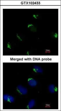
Bag1 antibody [N3C3]GTX102433
ApplicationsImmunoFluorescence, Western Blot, ImmunoCytoChemistry
ReactivityHuman
TargetBAG1
- SizePrice
Product group Antibodies
Anti-BAG1 AntibodyHPA018121
ApplicationsImmunoHistoChemistry
ReactivityHuman
TargetBAG1
- SizePrice
Product group Antibodies
Anti-BAG1 AntibodyCAB1104
ApplicationsImmunoFluorescence, Western Blot, ELISA, ImmunoCytoChemistry
ReactivityHuman
TargetBAG1
- SizePrice