

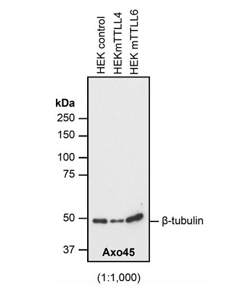
Figure 1: Immunoblot analysis of beta-tubulin protein glutamylation using anti-beta-Tubulin, mAb (AXO45) (Prod. No. AG-20B-0085). Method: HEK-293T cells are grown in standard culture conditions, transfected with plasmids expressing the different glutamylases, TTLL4 and TTLL6 and are run on a 10% SDS-PAGE (specific protocol to separate alpha- and beta-tubulin (Magiera & Janke, 2013)). The proteins are transferred to a nitrocellulose membrane and detected by standard immunoblot protocol using the AXO45 (1:1000) in PBS containing 0.1% Tween-20 for washing steps and 2.5% fat free milk for antibody incubation. In all the cell extracts, AXO45 specifically detects beta-tubulin, with no cross-reactivity to alpha-tubulin. Glutamylation added by either TTLL4 or TTLL6 appears to not affect this detection. Picture courtesy of Sudarshan Gadadhar, Shreyangi Chakraborty, Mariya Genova and Carsten Janke, Institut Curie, Orsay.
Figure 1: Immunoblot analysis of beta-tubulin protein glutamylation using anti-beta-Tubulin, mAb (AXO45) (Prod. No. AG-20B-0085). Method: HEK-293T cells are grown in standard culture conditions, transfected with plasmids expressing the different glutamylases, TTLL4 and TTLL6 and are run on a 10% SDS-PAGE (specific protocol to separate alpha- and beta-tubulin (Magiera & Janke, 2013)). The proteins are transferred to a nitrocellulose membrane and detected by standard immunoblot protocol using the AXO45 (1:1000) in PBS containing 0.1% Tween-20 for washing steps and 2.5% fat free milk for antibody incubation. In all the cell extracts, AXO45 specifically detects beta-tubulin, with no cross-reactivity to alpha-tubulin. Glutamylation added by either TTLL4 or TTLL6 appears to not affect this detection. Picture courtesy of Sudarshan Gadadhar, Shreyangi Chakraborty, Mariya Genova and Carsten Janke, Institut Curie, Orsay.
anti-beta-Tubulin, mAb (AXO45)
AG-20B-0085
ApplicationsImmunoPrecipitation, Western Blot, ImmunoCytoChemistry, ImmunoHistoChemistry
Product group Antibodies
ReactivityHuman, Mouse, Other Species
Overview
- SupplierAdipoGen Life Sciences
- Product Nameanti-beta-Tubulin, mAb (AXO45)
- Delivery Days Customer10
- ApplicationsImmunoPrecipitation, Western Blot, ImmunoCytoChemistry, ImmunoHistoChemistry
- CertificationResearch Use Only
- ClonalityMonoclonal
- Clone IDAXO45
- Concentration1 mg/ml
- Estimated Purity>95%
- HostMouse
- IsotypeIgG1
- Scientific DescriptionMonoclonal Antibody. Detects almost all isotypes of beta-tubulin specifically in cells and tissues of mouse, human and ciliates. Applications: ICC, IHC, IP and WB. Clone: AXO45. Isotype: Mouse IgG1. The beta-tubulin monoclonal antibody (AXO45) was developed by isolating the cilia from unicellular multiciliated Paramecium and purifying the core microtubule-based structures of the cilia, the axonemes. These axonemes were electrophoresed on a SDS-PAGE and the tubulin band was used as an antigen to develop monoclonal antibodies targeting both alpha- and beta-tubulin (A.M. Callen, et al.; 1994). The antibody AXO45 recognizes the C-terminal region between residues 257 and 442 of beta-tubulin. Although the antibody was raised against the beta-tubulin found in cilia, it is able to recognize beta-tubulin of all microtubule networks, indicating that it can potentially recognize different isotypes of beta-tubulin found in mammals. - The beta-tubulin monoclonal antibody (AXO45) was developed by isolating the cilia from unicellular multiciliated Paramecium and purifying the core microtubule-based structures of the cilia, the axonemes. These axonemes were electrophoresed on a SDS-PAGE and the tubulin band was used as an antigen to develop monoclonal antibodies targeting both alpha- and beta-tubulin (A.M. Callen, et al.; 1994). The antibody AXO45 recognizes the C-terminal region between residues 257 and 442 of beta-tubulin. Although the antibody was raised against the beta-tubulin found in cilia, it is able to recognize beta-tubulin of all microtubule networks, indicating that it can potentially recognize different isotypes of beta-tubulin found in mammals.
- ReactivityHuman, Mouse, Other Species
- Storage Instruction-20°C,2°C to 8°C
- UNSPSC41116161
MSDS
Related products
Product group Antibodies
anti-beta-Tubulin (beta-monoE), pAb (IN115)AG-25B-0039
ApplicationsImmunoPrecipitation, Western Blot, ImmunoCytoChemistry, ImmunoHistoChemistry
ReactivityHuman, Mouse
TargetTUBB2A
- SizePrice
Product group Antibodies
anti-beta-Tubulin, mAb (rec.) (S11B)AG-27B-0008
ApplicationsWestern Blot, ELISA, ImmunoCytoChemistry
ReactivityDrosophila, Human, Monkey, Mouse, Porcine, Rat
- SizePrice