


Figure 1. Western blot analysis of BEX3 using anti-BEX3 antibody (A09675-2). Electrophoresis was performed on a 5-20% SDS-PAGE gel at 70V (Stacking gel) / 90V (Resolving gel) for 2-3 hours. The sample well of each lane was loaded with 30 ug of sample under reducing conditions. Lane 1: human HCCT tissue lysates, Lane 2: human HCCP tissue lysates, Lane 3: human MCF-7 whole cell lysates, Lane 4: human Hela whole cell lysates, Lane 5: rat liver tissue lysates, Lane 6: rat L6 whole cell lysates, Lane 7: mouse liver tissue lysates, Lane 8: mouse C2C12 whole cell lysates. After electrophoresis, proteins were transferred to a nitrocellulose membrane at 150 mA for 50-90 minutes. Blocked the membrane with 5% non-fat milk/TBS for 1.5 hour at RT. The membrane was incubated with rabbit anti-BEX3 antigen affinity purified polyclonal antibody (Catalog # A09675-2) at 0.5 microg/mL overnight at 4°C, then washed with TBS-0.1%Tween 3 times with 5 minutes each and probed with a goat anti-rabbit IgG-HRP secondary antibody at a dilution of 1:5000 for 1.5 hour at RT. The signal is developed using an Enhanced Chemiluminescent detection (ECL) kit (Catalog # EK1002) with Tanon 5200 system. A specific band was detected for BEX3 at approximately 28 kDa. The expected band size for BEX3 is at 28 kDa.
. Overlay histogram showing MCF-7 cells stained with A09675-2 (Blue line). The cells were blocked with 10% normal goat serum. And then incubated with rabbit anti-BEX3 Antibody (A09675-2, 1 microg/1x106 cells) for 30 min at 20°C. DyLight?488 conjugated goat anti-rabbit IgG (BA1127, 5-10 microg/1x106 cells) was used as secondary antibody for 30 minutes at 20°C. Isotype control antibody (Green line) was rabbit IgG (1 microg/1x106) used under the same conditions. Unlabelled sample (Red line) was also used as a control.")
Figure 1. Western blot analysis of BEX3 using anti-BEX3 antibody (A09675-2). Electrophoresis was performed on a 5-20% SDS-PAGE gel at 70V (Stacking gel) / 90V (Resolving gel) for 2-3 hours. The sample well of each lane was loaded with 30 ug of sample under reducing conditions. Lane 1: human HCCT tissue lysates, Lane 2: human HCCP tissue lysates, Lane 3: human MCF-7 whole cell lysates, Lane 4: human Hela whole cell lysates, Lane 5: rat liver tissue lysates, Lane 6: rat L6 whole cell lysates, Lane 7: mouse liver tissue lysates, Lane 8: mouse C2C12 whole cell lysates. After electrophoresis, proteins were transferred to a nitrocellulose membrane at 150 mA for 50-90 minutes. Blocked the membrane with 5% non-fat milk/TBS for 1.5 hour at RT. The membrane was incubated with rabbit anti-BEX3 antigen affinity purified polyclonal antibody (Catalog # A09675-2) at 0.5 microg/mL overnight at 4°C, then washed with TBS-0.1%Tween 3 times with 5 minutes each and probed with a goat anti-rabbit IgG-HRP secondary antibody at a dilution of 1:5000 for 1.5 hour at RT. The signal is developed using an Enhanced Chemiluminescent detection (ECL) kit (Catalog # EK1002) with Tanon 5200 system. A specific band was detected for BEX3 at approximately 28 kDa. The expected band size for BEX3 is at 28 kDa.
Anti-BEX3 Antibody Picoband(r)
A09675-2-CARRIER-FREE
ApplicationsFlow Cytometry, Western Blot, ELISA
Product group Antibodies
ReactivityHuman, Mouse, Rat
TargetBEX3
Overview
- SupplierBoster Bio
- Product NameAnti-BEX3 Antibody Picoband(r)
- Delivery Days Customer9
- ApplicationsFlow Cytometry, Western Blot, ELISA
- CertificationResearch Use Only
- ClonalityPolyclonal
- Concentration500 ug/ml
- Gene ID27018
- Target nameBEX3
- Target descriptionbrain expressed X-linked 3
- Target synonymsBex, DXS6984E, HGR74, Hero20, NADE, NGFRAP1, protein BEX3, brain-expressed X-linked protein 3, nerve growth factor receptor (TNFRSF16) associated protein 1, ovarian granulosa cell 13.0 kDa protein HGR74, p75NTR-associated cell death executor
- HostRabbit
- IsotypeIgG
- Protein IDQ00994
- Protein NameProtein BEX3
- Scientific DescriptionBoster Bio Anti-BEX3 Antibody Picoband® catalog # A09675-2. Tested in ELISA, Flow Cytometry, WB applications. This antibody reacts with Human, Mouse, Rat. The brand Picoband indicates this is a premium antibody that guarantees superior quality, high affinity, and strong signals with minimal background in Western blot applications. Only our best-performing antibodies are designated as Picoband, ensuring unmatched performance.
- ReactivityHuman, Mouse, Rat
- Storage Instruction-20°C,2°C to 8°C
- UNSPSC12352203
Related products
Product group Antibodies
BEX3 AntibodyCSB-PA015781ESR1HU
ApplicationsELISA, ImmunoHistoChemistry
ReactivityHuman
TargetBEX3
- SizePrice
Product group Antibodies
Anti-NGFRAP1 AntibodyA31984
ApplicationsImmunoFluorescence, Western Blot, ImmunoHistoChemistry
ReactivityHuman
- SizePrice
Product group Antibodies

Goat anti-NGFRAP1EB07903
ApplicationsELISA
ReactivityHuman, Mouse, Rat
TargetBEX3
- SizePrice

Product group Antibodies
Anti-BEX3 AntibodyHPA018886
ApplicationsImmunoCytoChemistry, ImmunoHistoChemistry
ReactivityHuman
TargetBEX3
- SizePrice
Product group Antibodies

NGFRAP1 / NADE / Bex AntibodyLS-C346340
ApplicationsImmunoFluorescence, Western Blot, ImmunoHistoChemistry
ReactivityHuman
TargetBEX3
- SizePrice
Product group Antibodies
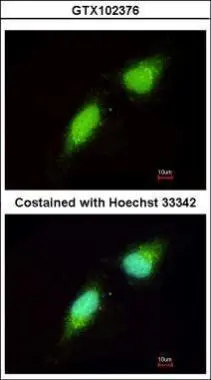
NADE antibody [N1C3]GTX102376
ApplicationsImmunoFluorescence, Western Blot, ImmunoCytoChemistry, ImmunoHistoChemistry, ImmunoHistoChemistry Paraffin
ReactivityHuman
TargetBEX3
- SizePrice
Product group Antibodies
Anti-NGFRAP1 Antibody144-07296
ApplicationsImmunoFluorescence, Western Blot
ReactivityHuman, Mouse
TargetBEX3
- SizePrice
Product group Antibodies
NGFRAP1 Polyclonal AntibodyBS-7671R
ApplicationsImmunoFluorescence, ELISA, ImmunoCytoChemistry, ImmunoHistoChemistry, ImmunoHistoChemistry Frozen, ImmunoHistoChemistry Paraffin
ReactivityBovine, Canine, Equine, Human, Mouse, Porcine, Rabbit, Rat
TargetBEX3
- SizePrice