


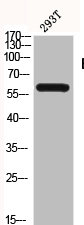
Figure 1. Western blot analysis of BLK using anti-BLK antibody (A01539-4). Electrophoresis was performed on a 5-20% SDS-PAGE gel at 70V (Stacking gel) / 90V (Resolving gel) for 2-3 hours. The sample well of each lane was loaded with 30 ug of sample under reducing conditions. Lane 1: human Daudi whole cell lysates. After electrophoresis, proteins were transferred to a nitrocellulose membrane at 150 mA for 50-90 minutes. Blocked the membrane with 5% non-fat milk/TBS for 1.5 hour at RT. The membrane was incubated with rabbit anti-BLK antigen affinity purified polyclonal antibody (Catalog # A01539-4) at 0.5 microg/mL overnight at 4°C, then washed with TBS-0.1%Tween 3 times with 5 minutes each and probed with a goat anti-rabbit IgG-HRP secondary antibody at a dilution of 1:5000 for 1.5 hour at RT. The signal is developed using an Enhanced Chemiluminescent detection (ECL) kit (Catalog # EK1002) with Tanon 5200 system. A specific band was detected for BLK at approximately 60 kDa. The expected band size for BLK is at 60 kDa.
. Overlay histogram showing Daudi cells stained with A01539-4 (Blue line). To facilitate intracellular staining, cells were fixed with 4% paraformaldehyde and permeabilized with permeabilization buffer. The cells were blocked with 10% normal goat serum. And then incubated with rabbit anti-BLK Antibody (A01539-4, 1 microg/1x106 cells) for 30 min at 20°C. DyLight®488 conjugated goat anti-rabbit IgG (BA1127, 5-10 microg/1x106 cells) was used as secondary antibody for 30 minutes at 20°C. Isotype control antibody (Green line) was rabbit IgG (1 microg/1x106) used under the same conditions. Unlabelled sample without incubation with primary antibody and secondary antibody (Red line) was used as a blank control.")
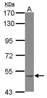
Figure 1. Western blot analysis of BLK using anti-BLK antibody (A01539-4). Electrophoresis was performed on a 5-20% SDS-PAGE gel at 70V (Stacking gel) / 90V (Resolving gel) for 2-3 hours. The sample well of each lane was loaded with 30 ug of sample under reducing conditions. Lane 1: human Daudi whole cell lysates. After electrophoresis, proteins were transferred to a nitrocellulose membrane at 150 mA for 50-90 minutes. Blocked the membrane with 5% non-fat milk/TBS for 1.5 hour at RT. The membrane was incubated with rabbit anti-BLK antigen affinity purified polyclonal antibody (Catalog # A01539-4) at 0.5 microg/mL overnight at 4°C, then washed with TBS-0.1%Tween 3 times with 5 minutes each and probed with a goat anti-rabbit IgG-HRP secondary antibody at a dilution of 1:5000 for 1.5 hour at RT. The signal is developed using an Enhanced Chemiluminescent detection (ECL) kit (Catalog # EK1002) with Tanon 5200 system. A specific band was detected for BLK at approximately 60 kDa. The expected band size for BLK is at 60 kDa.
Anti-BLK Antibody Picoband(r)
A01539-4-CARRIER-FREE
ApplicationsFlow Cytometry, Western Blot, ELISA
Product group Antibodies
ReactivityHuman
TargetBLK
Overview
- SupplierBoster Bio
- Product NameAnti-BLK Antibody Picoband(r)
- Delivery Days Customer9
- ApplicationsFlow Cytometry, Western Blot, ELISA
- CertificationResearch Use Only
- ClonalityPolyclonal
- Concentration500 ug/ml
- Gene ID640
- Target nameBLK
- Target descriptionBLK proto-oncogene, Src family tyrosine kinase
- Target synonymsMODY11, tyrosine-protein kinase Blk, B lymphoid tyrosine kinase, BLK nonreceptor tyrosine kinase, b lymphocyte kinase, p55-Blk
- HostRabbit
- IsotypeIgG
- Protein IDP51451
- Protein NameTyrosine-protein kinase Blk
- Scientific DescriptionBoster Bio Anti-BLK Antibody Picoband® catalog # A01539-4. Tested in ELISA, Flow Cytometry, WB applications. This antibody reacts with Human. The brand Picoband indicates this is a premium antibody that guarantees superior quality, high affinity, and strong signals with minimal background in Western blot applications. Only our best-performing antibodies are designated as Picoband, ensuring unmatched performance.
- ReactivityHuman
- Storage Instruction-20°C,2°C to 8°C
- UNSPSC12352203
Related products

Product group Antibodies
Anti-BLK AntibodyA98320
ApplicationsELISA, ImmunoHistoChemistry
ReactivityHuman, Mouse, Rat
- SizePrice
Product group Antibodies

BLK AntibodyLS-C760874
ApplicationsWestern Blot
ReactivityHuman, Mouse, Rat
TargetBLK
- SizePrice
Product group Antibodies
BLK Polyclonal AntibodyBS-6646R
ApplicationsImmunoFluorescence, Western Blot, ELISA, ImmunoCytoChemistry, ImmunoHistoChemistry, ImmunoHistoChemistry Frozen, ImmunoHistoChemistry Paraffin
ReactivityHuman, Mouse, Rat
TargetBLK
- SizePrice
Product group Antibodies
BLK AntibodyCSB-PA000997
ApplicationsWestern Blot, ELISA, ImmunoHistoChemistry
ReactivityHuman, Mouse, Rat
TargetBLK
- SizePrice
Product group Antibodies

Goat anti-BLKEB10736
ApplicationsWestern Blot, ELISA, ImmunoHistoChemistry
ReactivityHuman
TargetBLK
- SizePrice
Product group Antibodies
ApplicationsImmunoPrecipitation, Western Blot, ImmunoCytoChemistry, ImmunoHistoChemistry
ReactivityMouse, Porcine, Rat
TargetBLK
- SizePrice
Product group Antibodies
BLK antibody [N3C2], InternalGTX102348
ApplicationsWestern Blot
ReactivityHuman
TargetBLK
- SizePrice

Product group Antibodies
Anti-BLK AntibodyHPA069571
ApplicationsImmunoCytoChemistry
ReactivityHuman
TargetBLK
- SizePrice