


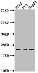
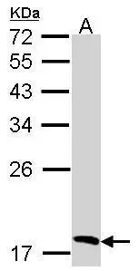
Figure 1. Western blot analysis of BLVRB using anti-BLVRB antibody (A08072-2). Electrophoresis was performed on a 5-20% SDS-PAGE gel at 70V (Stacking gel) / 90V (Resolving gel) for 2-3 hours. The sample well of each lane was loaded with 30 ug of sample under reducing conditions. Lane 1: human K562 whole cell lysates, Lane 2: human MCF-7 whole cell lysates, Lane 3: human A549 whole cell lysates, Lane 4: rat liver tissue lysates, Lane 5: rat RH35 whole cell lysates, Lane 6: mouse HEPA1-6 whole cell lysates. After electrophoresis, proteins were transferred to a nitrocellulose membrane at 150 mA for 50-90 minutes. Blocked the membrane with 5% non-fat milk/TBS for 1.5 hour at RT. The membrane was incubated with rabbit anti-BLVRB antigen affinity purified polyclonal antibody (Catalog # A08072-2) at 0.5 microg/mL overnight at 4°C, then washed with TBS-0.1%Tween 3 times with 5 minutes each and probed with a goat anti-rabbit IgG-HRP secondary antibody at a dilution of 1:5000 for 1.5 hour at RT. The signal is developed using an Enhanced Chemiluminescent detection (ECL) kit (Catalog # EK1002) with Tanon 5200 system. A specific band was detected for BLVRB at approximately 22 kDa. The expected band size for BLVRB is at 22-27 kDa.
. BLVRB was detected in an immunocytochemical section of A549 cells. Enzyme antigen retrieval was performed using IHC enzyme antigen retrieval reagent (AR0022) for 15 mins. The cells were blocked with 10% goat serum. And then incubated with 5 microg/mL rabbit anti-BLVRB Antibody (A08072-2) overnight at 4°C. Cy3 Conjugated Goat Anti-Rabbit IgG (BA1032) was used as secondary antibody at 1:500 dilution and incubated for 30 minutes at 37°C. The section was counterstained with DAPI. Visualize using a fluorescence microscope and filter sets appropriate for the label used.")
. Overlay histogram showing MCF-7 cells stained with A08072-2 (Blue line). To facilitate intracellular staining, cells were fixed with 4% paraformaldehyde and permeabilized with permeabilization buffer. The cells were blocked with 10% normal goat serum. And then incubated with rabbit anti-BLVRB Antibody (A08072-2, 1 microg/1x106 cells) for 30 min at 20°C. DyLight®488 conjugated goat anti-rabbit IgG (BA1127, 5-10 microg/1x106 cells) was used as secondary antibody for 30 minutes at 20°C. Isotype control antibody (Green line) was rabbit IgG (1 microg/1x106) used under the same conditions. Unlabelled sample (Red line) was also used as a control.")
Figure 1. Western blot analysis of BLVRB using anti-BLVRB antibody (A08072-2). Electrophoresis was performed on a 5-20% SDS-PAGE gel at 70V (Stacking gel) / 90V (Resolving gel) for 2-3 hours. The sample well of each lane was loaded with 30 ug of sample under reducing conditions. Lane 1: human K562 whole cell lysates, Lane 2: human MCF-7 whole cell lysates, Lane 3: human A549 whole cell lysates, Lane 4: rat liver tissue lysates, Lane 5: rat RH35 whole cell lysates, Lane 6: mouse HEPA1-6 whole cell lysates. After electrophoresis, proteins were transferred to a nitrocellulose membrane at 150 mA for 50-90 minutes. Blocked the membrane with 5% non-fat milk/TBS for 1.5 hour at RT. The membrane was incubated with rabbit anti-BLVRB antigen affinity purified polyclonal antibody (Catalog # A08072-2) at 0.5 microg/mL overnight at 4°C, then washed with TBS-0.1%Tween 3 times with 5 minutes each and probed with a goat anti-rabbit IgG-HRP secondary antibody at a dilution of 1:5000 for 1.5 hour at RT. The signal is developed using an Enhanced Chemiluminescent detection (ECL) kit (Catalog # EK1002) with Tanon 5200 system. A specific band was detected for BLVRB at approximately 22 kDa. The expected band size for BLVRB is at 22-27 kDa.
Anti-BLVRB Antibody Picoband(r)
A08072-2-CARRIER-FREE
ApplicationsFlow Cytometry, ImmunoFluorescence, Western Blot, ELISA, ImmunoCytoChemistry
Product group Antibodies
ReactivityHuman, Mouse, Rat
TargetBLVRB
Overview
- SupplierBoster Bio
- Product NameAnti-BLVRB Antibody Picoband(r)
- Delivery Days Customer9
- ApplicationsFlow Cytometry, ImmunoFluorescence, Western Blot, ELISA, ImmunoCytoChemistry
- CertificationResearch Use Only
- ClonalityPolyclonal
- Concentration500 ug/ml
- Gene ID645
- Target nameBLVRB
- Target descriptionbiliverdin reductase B
- Target synonymsBVRB, FLR, HEL-S-10, SDR43U1, flavin reductase (NADPH), BVR-B, FR, GHBP, NADPH-dependent diaphorase, NADPH-flavin reductase, S-nitroso-CoA-assisted nitrosyltransferase, SNO-CoA-assisted nitrosyltransferase, biliverdin-IX beta-reductase, epididymis secretory protein Li 10, green heme-binding protein, short chain dehydrogenase/reductase family 43U, member 1
- HostRabbit
- IsotypeIgG
- Protein IDP30043
- Protein NameFlavin reductase (NADPH)
- Scientific DescriptionBoster Bio Anti-BLVRB Antibody Picoband® catalog # A08072-2. Tested in ELISA, IF, ICC, WB, Flow Cytometry applications. This antibody reacts with Human, Mouse, Rat. The brand Picoband indicates this is a premium antibody that guarantees superior quality, high affinity, and strong signals with minimal background in Western blot applications. Only our best-performing antibodies are designated as Picoband, ensuring unmatched performance.
- ReactivityHuman, Mouse, Rat
- Storage Instruction-20°C,2°C to 8°C
- UNSPSC12352203
Related products
Product group Antibodies
Anti-BLVRB Antibody144-07260
ApplicationsWestern Blot, ImmunoHistoChemistry
ReactivityHuman, Mouse, Rat
TargetBLVRB
- SizePrice
Product group Antibodies
Anti-BLVRB AntibodyA31962
ApplicationsWestern Blot, ImmunoHistoChemistry
ReactivityHuman
- SizePrice
Product group Antibodies
BLVRB Polyclonal AntibodyCAC15771
ApplicationsImmunoFluorescence, Western Blot, ELISA, ImmunoHistoChemistry
TargetBLVRB
- SizePrice
Product group Antibodies
BLVRB AntibodyCSB-PA002722HA01HU
ApplicationsImmunoFluorescence, Western Blot, ELISA, ImmunoHistoChemistry
ReactivityHuman
TargetBLVRB
- SizePrice
Product group Antibodies
ApplicationsWestern Blot
ReactivityHuman
TargetBLVRB
- SizePrice

Product group Antibodies
Anti-BLVRB AntibodyHPA041698
ApplicationsWestern Blot, ImmunoCytoChemistry, ImmunoHistoChemistry
ReactivityHuman
TargetBLVRB
- SizePrice
Product group Antibodies

Goat anti-BLVRBEB12400
ApplicationsWestern Blot, ELISA
ReactivityHuman
TargetBLVRB
- SizePrice
Product group Antibodies

GHBP / BLVRB AntibodyLS-C831159
ApplicationsWestern Blot, ELISA, ImmunoHistoChemistry
ReactivityHuman, Mouse
TargetBLVRB
- SizePrice
Product group Antibodies
Anti-BLVRB AntibodyCAB7260
ApplicationsWestern Blot, ELISA, ImmunoHistoChemistry, ImmunoHistoChemistry Paraffin
ReactivityHuman
- SizePrice