

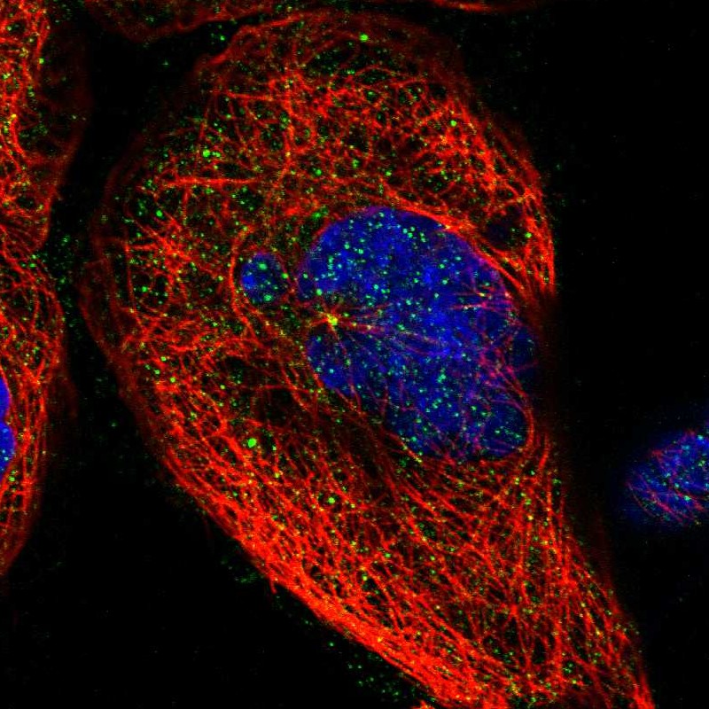
Immunofluorescent staining of human cell line A-431 shows localization to centrosome.

Immunofluorescent staining of human cell line A-431 shows localization to centrosome.
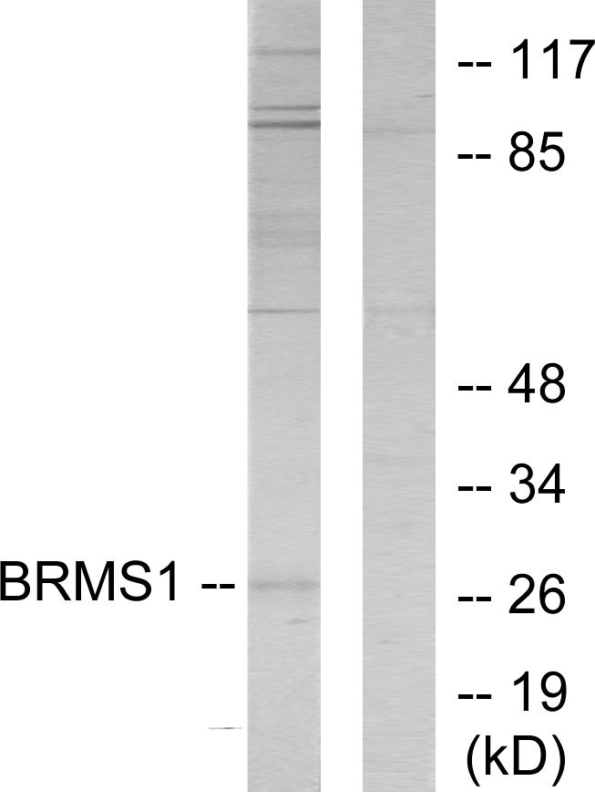
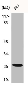
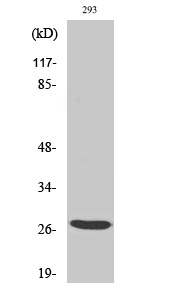
Anti-BRMS1 Antibody
HPA019637
ApplicationsWestern Blot, ImmunoCytoChemistry
Product group Antibodies
ReactivityHuman
TargetBRMS1
Overview
- SupplierAtlas Antibodies
- Product NameAnti-BRMS1 Antibody
- Delivery Days Customer4
- ApplicationsWestern Blot, ImmunoCytoChemistry
- CertificationResearch Use Only
- ClonalityPolyclonal
- ConjugateUnconjugated
- Gene ID25855
- Target nameBRMS1
- Target descriptionBRMS1 transcriptional repressor and anoikis regulator
- Target synonymsbreast cancer metastasis-suppressor 1, breast cancer metastasis suppressor 1
- HostRabbit
- IsotypeIgG
- Protein IDQ9HCU9
- Protein NameBreast cancer metastasis-suppressor 1
- Scientific DescriptionRecombinant Protein Epitope Signature Tag (PrEST) antigen sequence
- ReactivityHuman
- Storage Instruction-20°C,2°C to 8°C
- UNSPSC41116161
Datasheet
MSDS
Related products
Product group Antibodies
Anti-BRMS1 AntibodyA97677
ApplicationsWestern Blot, ELISA
ReactivityHuman, Mouse, Rat
- SizePrice

Product group Antibodies
Anti-BRMS1 Antibody Picoband(r)A03587-1-CARRIER-FREE
ApplicationsWestern Blot
ReactivityHuman, Mouse, Rat
TargetBRMS1
- SizePrice
Product group Antibodies
Anti-BRMS1 Antibody144-60829
ApplicationsWestern Blot, ImmunoHistoChemistry
ReactivityHuman, Mouse, Rat
TargetBRMS1
- SizePrice
Product group Antibodies

BRMS1 AntibodyLS-C749844
ApplicationsWestern Blot
ReactivityHuman, Mouse
TargetBRMS1
- SizePrice
Product group Antibodies
BRMS1 Recombinant Antibody, AbBy Fluor-405 ConjugatedBSM-62467R-BF405
ApplicationsImmunoFluorescence
ReactivityHuman
TargetBRMS1
- SizePrice
Product group Antibodies
BRMS1 AntibodyCSB-PA001048
ApplicationsWestern Blot, ELISA, ImmunoHistoChemistry
ReactivityHuman, Mouse, Rat
TargetBRMS1
- SizePrice
Product group Antibodies
BRMS1 antibodyGTX34063
ApplicationsWestern Blot
ReactivityHuman
TargetBRMS1
- SizePrice
Product group Antibodies
ApplicationsWestern Blot, ELISA, ImmunoHistoChemistry, ImmunoHistoChemistry Paraffin
ReactivityHuman
TargetBRMS1
- SizePrice