


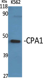
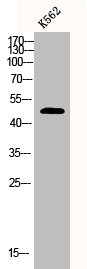
Figure 1. Western blot analysis of Carboxypeptidase A/CPA1 using anti-Carboxypeptidase A/CPA1 antibody (A05985-1). Electrophoresis was performed on a 5-20% SDS-PAGE gel at 70V (Stacking gel) / 90V (Resolving gel) for 2-3 hours. The sample well of each lane was loaded with 30 ug of sample under reducing conditions. Lane 1: rat pancreas tissue lysates, Lane 2: mouse pancreas tissue lysates. After electrophoresis, proteins were transferred to a nitrocellulose membrane at 150 mA for 50-90 minutes. Blocked the membrane with 5% non-fat milk/TBS for 1.5 hour at RT. The membrane was incubated with rabbit anti-Carboxypeptidase A/CPA1 antigen affinity purified polyclonal antibody (Catalog # A05985-1) at 0.5 microg/mL overnight at 4°C, then washed with TBS-0.1%Tween 3 times with 5 minutes each and probed with a goat anti-rabbit IgG-HRP secondary antibody at a dilution of 1:5000 for 1.5 hour at RT. The signal is developed using an Enhanced Chemiluminescent detection (ECL) kit (Catalog # EK1002) with Tanon 5200 system. A specific band was detected for Carboxypeptidase A/CPA1 at approximately 47 kDa. The expected band size for Carboxypeptidase A/CPA1 is at 47 kDa.
Figure 1. Western blot analysis of Carboxypeptidase A/CPA1 using anti-Carboxypeptidase A/CPA1 antibody (A05985-1). Electrophoresis was performed on a 5-20% SDS-PAGE gel at 70V (Stacking gel) / 90V (Resolving gel) for 2-3 hours. The sample well of each lane was loaded with 30 ug of sample under reducing conditions. Lane 1: rat pancreas tissue lysates, Lane 2: mouse pancreas tissue lysates. After electrophoresis, proteins were transferred to a nitrocellulose membrane at 150 mA for 50-90 minutes. Blocked the membrane with 5% non-fat milk/TBS for 1.5 hour at RT. The membrane was incubated with rabbit anti-Carboxypeptidase A/CPA1 antigen affinity purified polyclonal antibody (Catalog # A05985-1) at 0.5 microg/mL overnight at 4°C, then washed with TBS-0.1%Tween 3 times with 5 minutes each and probed with a goat anti-rabbit IgG-HRP secondary antibody at a dilution of 1:5000 for 1.5 hour at RT. The signal is developed using an Enhanced Chemiluminescent detection (ECL) kit (Catalog # EK1002) with Tanon 5200 system. A specific band was detected for Carboxypeptidase A/CPA1 at approximately 47 kDa. The expected band size for Carboxypeptidase A/CPA1 is at 47 kDa.
Anti-Carboxypeptidase A/CPA1 Antibody Picoband(r)
A05985-1-CARRIER-FREE
ApplicationsWestern Blot
Product group Antibodies
ReactivityMouse, Rat
TargetCPA1
Overview
- SupplierBoster Bio
- Product NameAnti-Carboxypeptidase A/CPA1 Antibody Picoband(r)
- Delivery Days Customer9
- ApplicationsWestern Blot
- CertificationResearch Use Only
- ClonalityPolyclonal
- Concentration500 ug/ml
- Gene ID1357
- Target nameCPA1
- Target descriptioncarboxypeptidase A1
- Target synonymsCPA, carboxypeptidase A1, carboxypeptidase A1 (pancreatic), pancreatic carboxypeptidase A
- HostRabbit
- IsotypeIgG
- Protein IDP15085
- Protein NameCarboxypeptidase A1
- Scientific DescriptionBoster Bio Anti-Carboxypeptidase A/CPA1 Antibody Picoband® catalog # A05985-1. Tested in WB applications. This antibody reacts with Mouse, Rat. The brand Picoband indicates this is a premium antibody that guarantees superior quality, high affinity, and strong signals with minimal background in Western blot applications. Only our best-performing antibodies are designated as Picoband, ensuring unmatched performance.
- ReactivityMouse, Rat
- Storage Instruction-20°C,2°C to 8°C
- UNSPSC12352203
Related products
Product group Antibodies
ApplicationsWestern Blot, ELISA, ImmunoHistoChemistry
ReactivityHuman
- SizePrice
Product group Antibodies
Anti-CPA1 Antibody144-03803
ApplicationsWestern Blot, ImmunoHistoChemistry
ReactivityHuman, Mouse, Rat
TargetCPA1
- SizePrice

Product group Antibodies
Anti-CPA1 AntibodyAMAB91433
ApplicationsWestern Blot, ImmunoHistoChemistry
ReactivityHuman
TargetCPA1
- SizePrice
Product group Antibodies

CPA1 / Carboxypeptidase A AntibodyLS-C748934
ApplicationsWestern Blot
ReactivityHuman, Mouse, Rat
TargetCPA1
- SizePrice
Product group Antibodies
ApplicationsImmunoFluorescence, Western Blot
ReactivityHuman, Mouse, Rat
TargetCPA1
- SizePrice
Product group Antibodies
CPA1 AntibodyCSB-PA001768
ApplicationsWestern Blot, ELISA, ImmunoHistoChemistry
ReactivityHuman
TargetCPA1
- SizePrice
![IHC-P analysis of human pancreas tissue using GTX04433 CPA1 antibody [MSVA-601M] HistoMAX?. Strong CPA1 immunostaining in acinar cells of the pancreas.](https://www.genetex.com/upload/website/prouct_img/normal/GTX04433/GTX04433_20230728_IHC-P_29_23072722_395.webp)
Product group Antibodies
ApplicationsImmunoHistoChemistry, ImmunoHistoChemistry Paraffin
ReactivityHuman
TargetCPA1
- SizePrice
Product group Antibodies
TargetCPA1
- SizePrice
Product group Antibodies
Anti-CPA1 AntibodyCAB3803
ApplicationsImmunoFluorescence, Western Blot, ELISA, ImmunoCytoChemistry
ReactivityMouse
TargetCPA1
- SizePrice