


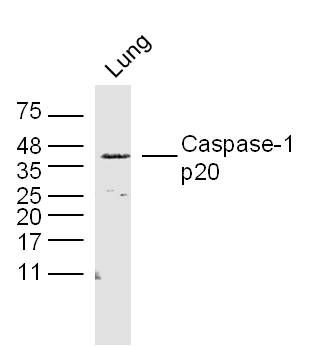
Figure 1. Western blot analysis of CASP1 using anti-CASP1 antibody (PA1440-1). Electrophoresis was performed on a 5-20% SDS-PAGE gel at 70V (Stacking gel) / 90V (Resolving gel) for 2-3 hours. The sample well of each lane was loaded with 30 ug of sample under reducing conditions. Lane 1: rat thymus tissue lysates, Lane 2: rat C6 whole cell lysates, Lane 3: mouse thymus tissue lysates. After electrophoresis, proteins were transferred to a nitrocellulose membrane at 150 mA for 50-90 minutes. Blocked the membrane with 5% non-fat milk/TBS for 1.5 hour at RT. The membrane was incubated with rabbit anti-CASP1 antigen affinity purified polyclonal antibody (Catalog # PA1440-1) at 0.5 microg/mL overnight at 4°C, then washed with TBS-0.1%Tween 3 times with 5 minutes each and probed with a goat anti-rabbit IgG-HRP secondary antibody at a dilution of 1:5000 for 1.5 hour at RT. The signal is developed using an Enhanced Chemiluminescent detection (ECL) kit (Catalog # EK1002) with Tanon 5200 system. A specific band was detected for CASP1 at approximately 46 kDa. The expected band size for CASP1 is at 46 kDa.
Figure 1. Western blot analysis of CASP1 using anti-CASP1 antibody (PA1440-1). Electrophoresis was performed on a 5-20% SDS-PAGE gel at 70V (Stacking gel) / 90V (Resolving gel) for 2-3 hours. The sample well of each lane was loaded with 30 ug of sample under reducing conditions. Lane 1: rat thymus tissue lysates, Lane 2: rat C6 whole cell lysates, Lane 3: mouse thymus tissue lysates. After electrophoresis, proteins were transferred to a nitrocellulose membrane at 150 mA for 50-90 minutes. Blocked the membrane with 5% non-fat milk/TBS for 1.5 hour at RT. The membrane was incubated with rabbit anti-CASP1 antigen affinity purified polyclonal antibody (Catalog # PA1440-1) at 0.5 microg/mL overnight at 4°C, then washed with TBS-0.1%Tween 3 times with 5 minutes each and probed with a goat anti-rabbit IgG-HRP secondary antibody at a dilution of 1:5000 for 1.5 hour at RT. The signal is developed using an Enhanced Chemiluminescent detection (ECL) kit (Catalog # EK1002) with Tanon 5200 system. A specific band was detected for CASP1 at approximately 46 kDa. The expected band size for CASP1 is at 46 kDa.
Anti-Caspase-1(P20)/CASP1 Antibody Picoband(r)
PA1440-1-CARRIER-FREE
ApplicationsWestern Blot
Product group Antibodies
ReactivityMouse, Rat
TargetCasp1
Overview
- SupplierBoster Bio
- Product NameAnti-Caspase-1(P20)/CASP1 Antibody Picoband(r)
- Delivery Days Customer9
- Application Supplier NoteTested Species: In-house tested species with positive results. Predicted Species: Species predicted to be fit for the product based on sequence similarities. By Heat: Boiling the paraffin sections in 10mM citrate buffer, pH6.0, for 20mins is required for the staining of formalin/paraffin sections. Other applications have not been tested. Optimal dilutions should be determined by end users.
- ApplicationsWestern Blot
- CertificationResearch Use Only
- ClonalityPolyclonal
- Concentration500 ug/ml
- Gene ID12362
- Target nameCasp1
- Target descriptioncaspase 1
- Target synonymsICE, Il1bc, caspase-1, CASP-1, IL-1 beta-converting enzyme, IL-1B converting enzyme, IL-1BC, interleukin-1 beta convertase, interleukin-1 beta-converting enzyme, p45
- HostRabbit
- IsotypeIgG
- Protein IDP29452
- Protein NameCaspase-1
- Scientific DescriptionBoster Bio Anti-Caspase-1 (P20)/CASP1 Antibody catalog # PA1440-1. Tested in WB applications. This antibody reacts with Mouse, Rat. The brand Picoband indicates this is a premium antibody that guarantees superior quality, high affinity, and strong signals with minimal background in Western blot applications. Only our best-performing antibodies are designated as Picoband, ensuring unmatched performance.
- ReactivityMouse, Rat
- Storage Instruction-20°C,2°C to 8°C
- UNSPSC12352203
Related products
Product group Antibodies
anti-Caspase-1 (p20) (mouse), mAb (Casper-1)AG-20B-0042
ApplicationsImmunoPrecipitation, Western Blot, ImmunoHistoChemistry
ReactivityMouse
TargetCasp1
- SizePrice
Product group Antibodies
Caspase-1 p20 Polyclonal AntibodyBS-10442R
ApplicationsFlow Cytometry, ImmunoFluorescence, Western Blot, ELISA, ImmunoCytoChemistry, ImmunoHistoChemistry, ImmunoHistoChemistry Frozen, ImmunoHistoChemistry Paraffin
ReactivityHuman, Mouse, Rat
TargetCasp1
- SizePrice
Product group Antibodies
ApplicationsImmunoPrecipitation, Western Blot, ImmunoCytoChemistry, ImmunoHistoChemistry
ReactivityMouse, Rat
TargetCasp1
- SizePrice
Product group Antibodies
References
Caspase 1 p20 subunit antibodyGTX11701
ApplicationsWestern Blot
ReactivityMouse, Rat
TargetCasp1
- SizePrice