


Figure 1. Western blot analysis of CCDC36 using anti-CCDC36 antibody (A14775). Electrophoresis was performed on a 5-20% SDS-PAGE gel at 70V (Stacking gel) / 90V (Resolving gel) for 2-3 hours. The sample well of each lane was loaded with 50ug of sample under reducing conditions. Lane 1: human placenta tissue lysates, Lane 2: human HL-60 whole cell lysates. After Electrophoresis, proteins were transferred to a Nitrocellulose membrane at 150mA for 50-90 minutes. Blocked the membrane with 5% Non-fat Milk/ TBS for 1.5 hour at RT. The membrane was incubated with rabbit anti-CCDC36 antigen affinity purified polyclonal antibody (Catalog # A14775) at 0.5 microg/mL overnight at 4°C, then washed with TBS-0.1%Tween 3 times with 5 minutes each and probed with a goat anti-rabbit IgG-HRP secondary antibody at a dilution of 1:10000 for 1.5 hour at RT. The signal is developed using an Enhanced Chemiluminescent detection (ECL) kit (Catalog # EK1002) with Tanon 5200 system. A specific band was detected for CCDC36 at approximately 66KD. The expected band size for CCDC36 is at 66KD.
. CCDC36 was detected in paraffin-embedded section of human testis cancer tissue. Heat mediated antigen retrieval was performed in citrate buffer (pH6, epitope retrieval solution) for 20 mins. The tissue section was blocked with 10% goat serum. The tissue section was then incubated with 1microg/ml rabbit anti-CCDC36 Antibody (A14775) overnight at 4°C. Biotinylated goat anti-rabbit IgG was used as secondary antibody and incubated for 30 minutes at 37°C. The tissue section was developed using Strepavidin-Biotin-Complex (SABC)(Catalog # SA1022) with DAB as the chromogen.")
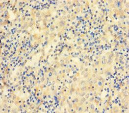
. CCDC36 was detected in paraffin-embedded section of human melanoma tissue. Heat mediated antigen retrieval was performed in citrate buffer (pH6, epitope retrieval solution) for 20 mins. The tissue section was blocked with 10% goat serum. The tissue section was then incubated with 1microg/ml rabbit anti-CCDC36 Antibody (A14775) overnight at 4°C. Biotinylated goat anti-rabbit IgG was used as secondary antibody and incubated for 30 minutes at 37°C. The tissue section was developed using Strepavidin-Biotin-Complex (SABC)(Catalog # SA1022) with DAB as the chromogen.")
. CCDC36 was detected in immunocytochemical section of SiHa cells. Enzyme antigen retrieval was performed using IHC enzyme antigen retrieval reagent (AR0022) for 15 mins. The cells were blocked with 10% goat serum. And then incubated with 2microg/mL rabbit anti-CCDC36 Antibody (A14775) overnight at 4°C. DyLight®488 Conjugated Goat Anti-Rabbit IgG (BA1127) was used as secondary antibody at 1:100 dilution and incubated for 30 minutes at 37°C. The section was counterstained with DAPI. Visualize using a fluorescence microscope and filter sets appropriate for the label used.")
Figure 1. Western blot analysis of CCDC36 using anti-CCDC36 antibody (A14775). Electrophoresis was performed on a 5-20% SDS-PAGE gel at 70V (Stacking gel) / 90V (Resolving gel) for 2-3 hours. The sample well of each lane was loaded with 50ug of sample under reducing conditions. Lane 1: human placenta tissue lysates, Lane 2: human HL-60 whole cell lysates. After Electrophoresis, proteins were transferred to a Nitrocellulose membrane at 150mA for 50-90 minutes. Blocked the membrane with 5% Non-fat Milk/ TBS for 1.5 hour at RT. The membrane was incubated with rabbit anti-CCDC36 antigen affinity purified polyclonal antibody (Catalog # A14775) at 0.5 microg/mL overnight at 4°C, then washed with TBS-0.1%Tween 3 times with 5 minutes each and probed with a goat anti-rabbit IgG-HRP secondary antibody at a dilution of 1:10000 for 1.5 hour at RT. The signal is developed using an Enhanced Chemiluminescent detection (ECL) kit (Catalog # EK1002) with Tanon 5200 system. A specific band was detected for CCDC36 at approximately 66KD. The expected band size for CCDC36 is at 66KD.
Anti-CCDC36 Antibody Picoband(r)
A14775-CARRIER-FREE
ApplicationsImmunoFluorescence, Western Blot, ELISA, ImmunoCytoChemistry, ImmunoHistoChemistry
Product group Antibodies
ReactivityHuman
TargetIHO1
Overview
- SupplierBoster Bio
- Product NameAnti-CCDC36 Antibody Picoband(r)
- Delivery Days Customer9
- Application Supplier NoteTested Species: In-house tested species with positive results. By Heat: Boiling the paraffin sections in 10mM citrate buffer, pH6.0, for 20mins is required for the staining of formalin/paraffin sections. Other applications have not been tested. Optimal dilutions should be determined by end users.
- ApplicationsImmunoFluorescence, Western Blot, ELISA, ImmunoCytoChemistry, ImmunoHistoChemistry
- CertificationResearch Use Only
- ClonalityPolyclonal
- Concentration500 ug/ml
- Gene ID339834
- Target nameIHO1
- Target descriptioninteractor of HORMAD1 1
- Target synonymsCCDC36, CT74, LELA1, interactor of HORMAD1 protein 1, cancer/testis antigen 74, coiled-coil domain containing 36, coiled-coil domain-containing protein 36
- HostRabbit
- IsotypeIgG
- Protein IDQ8IYA8
- Protein NameInteractor of HORMAD1 protein 1
- Scientific DescriptionBoster Bio Anti-CCDC36 Antibody Picoband® catalog # A14775. Tested in ELISA, IF, IHC, ICC, WB applications. This antibody reacts with Human. The brand Picoband indicates this is a premium antibody that guarantees superior quality, high affinity, and strong signals with minimal background in Western blot applications. Only our best-performing antibodies are designated as Picoband, ensuring unmatched performance.
- ReactivityHuman
- Storage Instruction-20°C,2°C to 8°C
- UNSPSC12352203
Related products
Product group Antibodies
CT74 Polyclonal AntibodyBS-8129R
ApplicationsImmunoFluorescence, Western Blot, ELISA, ImmunoCytoChemistry, ImmunoHistoChemistry, ImmunoHistoChemistry Frozen, ImmunoHistoChemistry Paraffin
ReactivityBovine, Equine, Human, Mouse, Rat, Sheep
TargetIHO1
- SizePrice
Product group Antibodies
CCDC36 AntibodyCSB-PA816910LA01HU
ApplicationsELISA, ImmunoHistoChemistry
ReactivityHuman
TargetIHO1
- SizePrice
Product group Antibodies

CCDC36 Antibody (aa11-592, HRP)LS-C371623
ApplicationsELISA
ReactivityHuman
TargetIHO1
- SizePrice

Product group Antibodies
Anti-CCDC36 AntibodyHPA045690
ApplicationsImmunoHistoChemistry
ReactivityHuman
TargetIHO1
- SizePrice
Product group Antibodies
CCDC36 AntibodyPACO37842
ApplicationsELISA, ImmunoHistoChemistry
ReactivityHuman
TargetIHO1
- SizePrice