


Figure 1. Western blot analysis of CCR4 using anti-CCR4 antibody (A00755-4). Electrophoresis was performed on a 5-20% SDS-PAGE gel at 70V (Stacking gel) / 90V (Resolving gel) for 2-3 hours. The sample well of each lane was loaded with 30 ug of sample under reducing conditions. Lane 1: human 293T whole cell lysates, Lane 2: human Raji whole cell lysates, Lane 3: rat spleen tissue lysates, Lane 4: rat thymus tissue lysates, Lane 5: mouse spleen tissue lysates, Lane 6: mouse thymus tissue lysates, Lane 7: mouse RAW264.7 whole cell lysates. After electrophoresis, proteins were transferred to a nitrocellulose membrane at 150 mA for 50-90 minutes. Blocked the membrane with 5% non-fat milk/TBS for 1.5 hour at RT. The membrane was incubated with rabbit anti-CCR4 antigen affinity purified polyclonal antibody (Catalog # A00755-4) at 0.5 microg/mL overnight at 4°C, then washed with TBS-0.1%Tween 3 times with 5 minutes each and probed with a goat anti-rabbit IgG-HRP secondary antibody at a dilution of 1:5000 for 1.5 hour at RT. The signal is developed using an Enhanced Chemiluminescent detection (ECL) kit (Catalog # EK1002) with Tanon 5200 system. A specific band was detected for CCR4 at approximately 50 kDa. The expected band size for CCR4 is at 41 kDa.
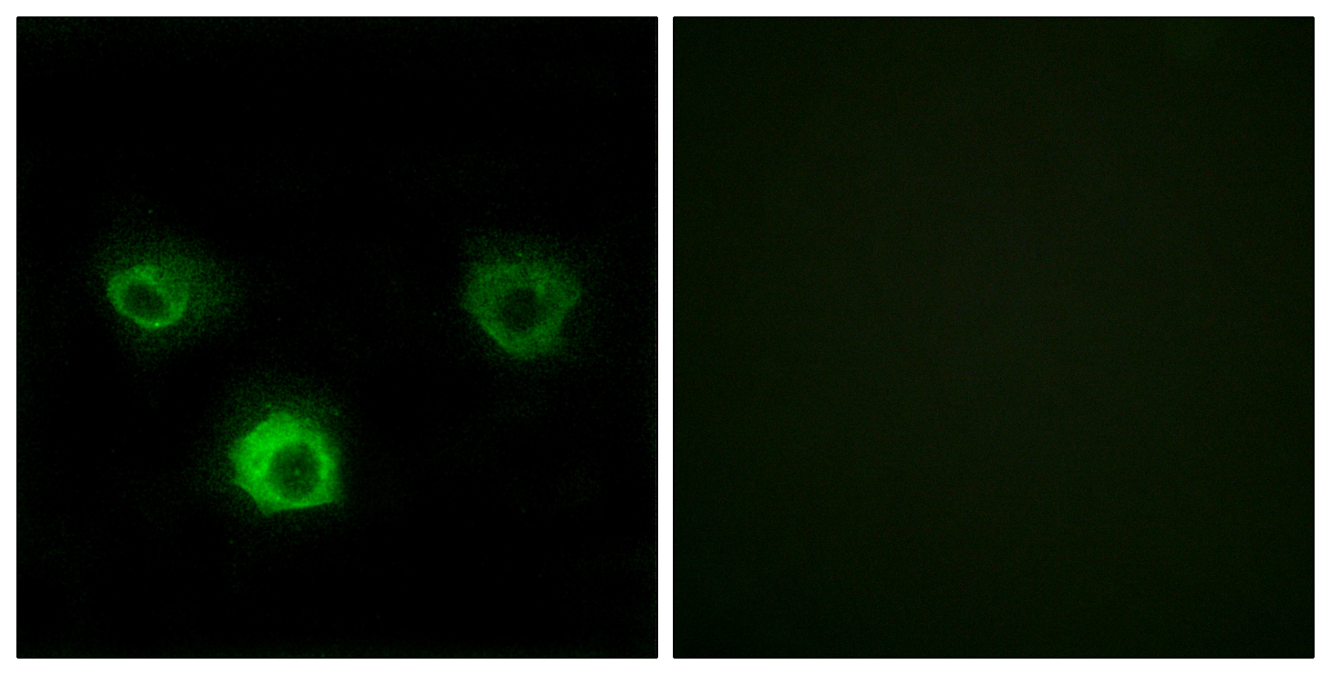
. Overlay histogram showing HEL cells stained with A00755-4 (Blue line). The cells were fixed with 4% paraformaldehyde and blocked with 10% normal goat serum. And then incubated with rabbit anti-CCR4 Antibody (A00755-4, 1 microg/1x106 cells) for 30 min at 20°C. DyLight®488 conjugated goat anti-rabbit IgG (BA1127, 5-10 microg/1x106 cells) was used as secondary antibody for 30 minutes at 20°C. Isotype control antibody (Green line) was rabbit IgG (1 microg/1x106) used under the same conditions. Unlabelled sample (Red line) was also used as a control.")
Figure 1. Western blot analysis of CCR4 using anti-CCR4 antibody (A00755-4). Electrophoresis was performed on a 5-20% SDS-PAGE gel at 70V (Stacking gel) / 90V (Resolving gel) for 2-3 hours. The sample well of each lane was loaded with 30 ug of sample under reducing conditions. Lane 1: human 293T whole cell lysates, Lane 2: human Raji whole cell lysates, Lane 3: rat spleen tissue lysates, Lane 4: rat thymus tissue lysates, Lane 5: mouse spleen tissue lysates, Lane 6: mouse thymus tissue lysates, Lane 7: mouse RAW264.7 whole cell lysates. After electrophoresis, proteins were transferred to a nitrocellulose membrane at 150 mA for 50-90 minutes. Blocked the membrane with 5% non-fat milk/TBS for 1.5 hour at RT. The membrane was incubated with rabbit anti-CCR4 antigen affinity purified polyclonal antibody (Catalog # A00755-4) at 0.5 microg/mL overnight at 4°C, then washed with TBS-0.1%Tween 3 times with 5 minutes each and probed with a goat anti-rabbit IgG-HRP secondary antibody at a dilution of 1:5000 for 1.5 hour at RT. The signal is developed using an Enhanced Chemiluminescent detection (ECL) kit (Catalog # EK1002) with Tanon 5200 system. A specific band was detected for CCR4 at approximately 50 kDa. The expected band size for CCR4 is at 41 kDa.
Anti-CCR4 Antibody Picoband(r)
A00755-4-CARRIER-FREE
ApplicationsFlow Cytometry, Western Blot, ELISA
Product group Antibodies
ReactivityHuman, Mouse, Rat
TargetCCR4
Overview
- SupplierBoster Bio
- Product NameAnti-CCR4 Antibody Picoband(r)
- Delivery Days Customer9
- ApplicationsFlow Cytometry, Western Blot, ELISA
- CertificationResearch Use Only
- ClonalityPolyclonal
- Concentration500 ug/ml
- Gene ID1233
- Target nameCCR4
- Target descriptionC-C motif chemokine receptor 4
- Target synonymsCC-CKR-4, CD194, CKR4, CMKBR4, ChemR13, HGCN:14099, K5-5, C-C chemokine receptor type 4, C-C CKR-4, CCR-4, chemokine (C-C motif) receptor 4, chemokine (C-C) receptor 4
- HostRabbit
- IsotypeIgG
- Protein IDP51679
- Protein NameC-C chemokine receptor type 4
- Scientific DescriptionBoster Bio Anti-CCR4 Antibody Picoband® catalog # A00755-4. Tested in ELISA, Flow Cytometry, WB applications. This antibody reacts with Human, Mouse, Rat. The brand Picoband indicates this is a premium antibody that guarantees superior quality, high affinity, and strong signals with minimal background in Western blot applications. Only our best-performing antibodies are designated as Picoband, ensuring unmatched performance.
- ReactivityHuman, Mouse, Rat
- Storage Instruction-20°C,2°C to 8°C
- UNSPSC12352203
Related products
Product group Antibodies
Anti-CCR4 AntibodyA98254
ApplicationsImmunoFluorescence, ELISA
ReactivityHuman, Mouse, Rat
- SizePrice
Product group Antibodies

Anti-CCR4 [KW-0761 (Mogamulizumab)]Ab00726-2.0
ApplicationsOther Application
ReactivityHuman
TargetCCR4
- SizePrice
Product group Antibodies
ApplicationsFlow Cytometry
ReactivityHuman
TargetCCR4
- SizePrice
Product group Antibodies
CCR4 Polyclonal AntibodyBS-1168R
ApplicationsImmunoFluorescence, ELISA, ImmunoCytoChemistry, ImmunoHistoChemistry, ImmunoHistoChemistry Frozen, ImmunoHistoChemistry Paraffin
ReactivityHuman, Mouse, Rat
TargetCCR4
- SizePrice
Product group Antibodies
Ccr4 Polyclonal AntibodyCAC11789
ApplicationsImmunoFluorescence, ELISA, ImmunoHistoChemistry
TargetCCR4
- SizePrice
Product group Antibodies
CCR4 AntibodyCSB-PA004843LA01HU
ApplicationsImmunoFluorescence, ELISA, ImmunoHistoChemistry
ReactivityHuman
TargetCCR4
- SizePrice
![Mouse tissue extract (50 μg) was separated by 7.5% SDS-PAGE, and the membrane was blotted with CCR4 antibody [HL2481] (GTX638830) diluted at 1:1000. The HRP-conjugated anti-rabbit IgG antibody (GTX213110-01) was used to detect the primary antibody.](https://www.genetex.com/upload/website/prouct_img/normal/GTX638830/GTX638830_T-45096_20230721_WB_M_thymus_23072519_504.webp)
Product group Antibodies
CCR4 antibody [HL2481]GTX638830
ApplicationsFlow Cytometry, ImmunoFluorescence, Western Blot, ImmunoCytoChemistry, ImmunoHistoChemistry, ImmunoHistoChemistry Paraffin
ReactivityHuman, Mouse, Rat
TargetCCR4
- SizePrice

Product group Antibodies
Anti-CCR4 AntibodyHPA031613
ApplicationsImmunoHistoChemistry
ReactivityHuman
TargetCCR4
- SizePrice
Product group Antibodies

CCR4 Antibody (HRP)LS-C396075
ApplicationsELISA
ReactivityHuman
TargetCCR4
- SizePrice