

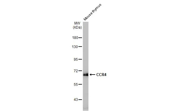
Mouse tissue extract (50 μg) was separated by 7.5% SDS-PAGE, and the membrane was blotted with CCR4 antibody [HL2481] (GTX638830) diluted at 1:1000. The HRP-conjugated anti-rabbit IgG antibody (GTX213110-01) was used to detect the primary antibody.
![Unboiled SW480 whole cell and membrane extracts (30 μg) were separated by 7.5% SDS-PAGE, and the membrane was blotted with CCR4 antibody [HL2481] (GTX638830) diluted at 1:500. The HRP-conjugated anti-rabbit IgG antibody (GTX213110-01) was used to detect the primary antibody, and the signal was developed with Trident ECL plus-Enhanced. (WCE: whole cell extract; ME: membrane extract)](https://www.genetex.com/upload/website/prouct_img/normal/GTX638830/GTX638830_T-45096_20230728_WB_Fraction_23073119_795.webp "Unboiled SW480 whole cell and membrane extracts (30 μg) were separated by 7.5% SDS-PAGE, and the membrane was blotted with CCR4 antibody [HL2481] (GTX638830) diluted at 1:500. The HRP-conjugated anti-rabbit IgG antibody (GTX213110-01) was used to detect the primary antibody, and the signal was developed with Trident ECL plus-Enhanced. (WCE: whole cell extract; ME: membrane extract)")
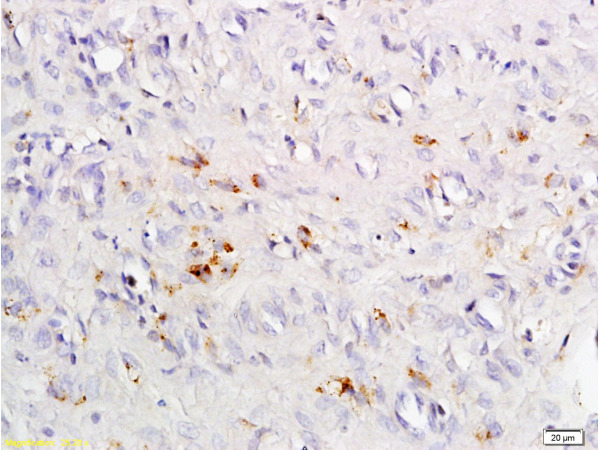
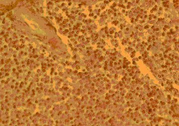
![CCR4 antibody [HL2481] detects CCR4 protein at cell membrane by immunohistochemical analysis. Sample: Paraffin-embedded mouse thymus gland. CCR4 stained by CCR4 antibody [HL2481] (GTX638830) diluted at 1:100. Antigen Retrieval: Citrate buffer, pH 6.0, 15 min](https://www.genetex.com/upload/website/prouct_img/normal/GTX638830/GTX638830_T-45096_20230721_IHC-P_M_23073119_736.webp "CCR4 antibody [HL2481] detects CCR4 protein at cell membrane by immunohistochemical analysis. Sample: Paraffin-embedded mouse thymus gland. CCR4 stained by CCR4 antibody [HL2481] (GTX638830) diluted at 1:100. Antigen Retrieval: Citrate buffer, pH 6.0, 15 min")
![CCR4 antibody [HL2481] detects CCR4 protein at cell membrane by immunohistochemical analysis. Sample: Paraffin-embedded rat thymus gland. CCR4 stained by CCR4 antibody [HL2481] (GTX638830) diluted at 1:100. Antigen Retrieval: Citrate buffer, pH 6.0, 15 min](https://www.genetex.com/upload/website/prouct_img/normal/GTX638830/GTX638830_T-45096_20230721_IHC-P_R_23073119_973.webp "CCR4 antibody [HL2481] detects CCR4 protein at cell membrane by immunohistochemical analysis. Sample: Paraffin-embedded rat thymus gland. CCR4 stained by CCR4 antibody [HL2481] (GTX638830) diluted at 1:100. Antigen Retrieval: Citrate buffer, pH 6.0, 15 min")
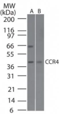
![Non-transfected (–) and transfected (+) boiled and unboiled SW480 whole cell extracts (30 μg) were separated by 10% SDS-PAGE, and the membrane was blotted with CCR4 antibody [HL2481] (GTX638830) diluted at 1:1000. The HRP-conjugated anti-rabbit IgG antibody (GTX213110-01) was used to detect the primary antibody.](https://www.genetex.com/upload/website/prouct_img/normal/GTX638830/GTX638830_T-45096_20230818_WB_B_23082201_891.webp "Non-transfected (–) and transfected (+) boiled and unboiled SW480 whole cell extracts (30 μg) were separated by 10% SDS-PAGE, and the membrane was blotted with CCR4 antibody [HL2481] (GTX638830) diluted at 1:1000. The HRP-conjugated anti-rabbit IgG antibody (GTX213110-01) was used to detect the primary antibody.")
![Non-transfected (–) and transfected (+) bolied and unboiled 293T whole cell extracts (30 μg) were separated by 10% SDS-PAGE, and the membrane was blotted with CCR4 antibody [HL2481] (GTX638830) diluted at 1:1000. The HRP-conjugated anti-rabbit IgG antibody (GTX213110-01) was used to detect the primary antibody.](https://www.genetex.com/upload/website/prouct_img/normal/GTX638830/GTX638830_45173_20230922_WB_B_23100118_107.webp "Non-transfected (–) and transfected (+) bolied and unboiled 293T whole cell extracts (30 μg) were separated by 10% SDS-PAGE, and the membrane was blotted with CCR4 antibody [HL2481] (GTX638830) diluted at 1:1000. The HRP-conjugated anti-rabbit IgG antibody (GTX213110-01) was used to detect the primary antibody.")
![Non-transfected (–) and transfected (+) boiled and unboiled 293T whole cell extracts (30 μg) were separated by 10% SDS-PAGE, and the membranes were blotted with CCR4 antibody [HL2481] (GTX638830) diluted at 1:1000 and the highly cited competitor's diluted at 1:1000. The HRP-conjugated anti-rabbit IgG antibody (GTX213110-01) was used to detect the primary antibody. *The competitor is not affiliated with GeneTex and does not endorse this product.](https://www.genetex.com/upload/website/prouct_img/normal/GTX638830/GTX638830_45173_20240301_WB_B_competitor_watermark_24030600_872.webp "Non-transfected (–) and transfected (+) boiled and unboiled 293T whole cell extracts (30 μg) were separated by 10% SDS-PAGE, and the membranes were blotted with CCR4 antibody [HL2481] (GTX638830) diluted at 1:1000 and the highly cited competitor's diluted at 1:1000. The HRP-conjugated anti-rabbit IgG antibody (GTX213110-01) was used to detect the primary antibody. *The competitor is not affiliated with GeneTex and does not endorse this product.")
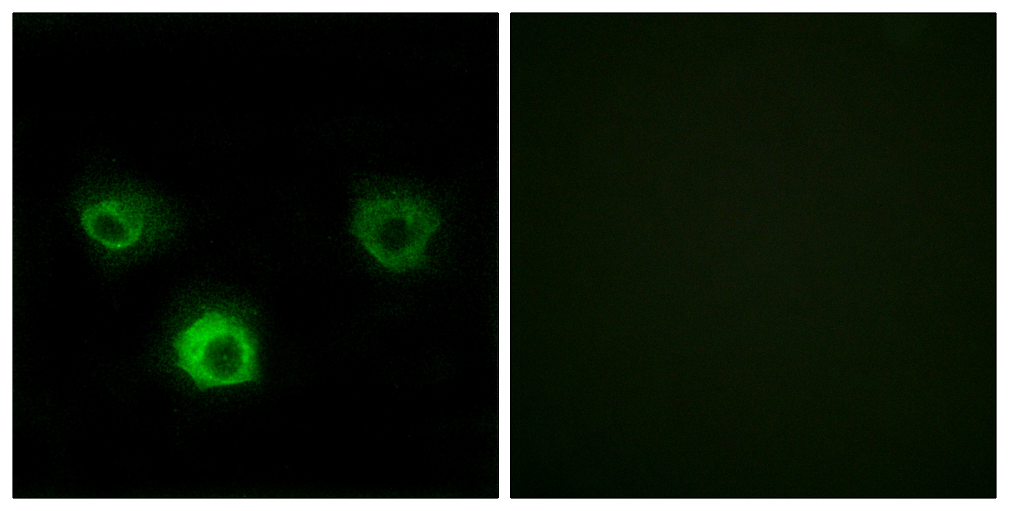
![CCR4 antibody [HL2481] detects CCR4 protein by immunofluorescent analysis. Sample: SW480 cells were fixed in ice-cold MeOH for 5 min. Green: CCR4 stained by CCR4 antibody [HL2481] (GTX638830) diluted at 1:200. Blue: Fluoroshield with DAPI (GTX30920).](https://www.genetex.com/upload/website/prouct_img/normal/GTX638830/GTX638830_45173_20240830_ICC_IF_KO_24091102_409.webp "CCR4 antibody [HL2481] detects CCR4 protein by immunofluorescent analysis. Sample: SW480 cells were fixed in ice-cold MeOH for 5 min. Green: CCR4 stained by CCR4 antibody [HL2481] (GTX638830) diluted at 1:200. Blue: Fluoroshield with DAPI (GTX30920).")
![CCR4 antibody [HL2481] detects CCR4 protein by immunohistochemical analysis. Sample: Paraffin-embedded human tonsil. CCR4 stained by CCR4 antibody [HL2481] (GTX638830) diluted at 1:500. Antigen Retrieval: Citrate buffer, pH 6.0, 15 min](https://www.genetex.com/upload/website/prouct_img/normal/GTX638830/GTX638830_T-44977_20241108_IHC-P_24111918_179.webp "CCR4 antibody [HL2481] detects CCR4 protein by immunohistochemical analysis. Sample: Paraffin-embedded human tonsil. CCR4 stained by CCR4 antibody [HL2481] (GTX638830) diluted at 1:500. Antigen Retrieval: Citrate buffer, pH 6.0, 15 min")
![CCR4 antibody [HL2481] detects CCR4 protein by immunohistochemical analysis. Sample: Paraffin-embedded mouse thymus gland. CCR4 stained by CCR4 antibody [HL2481] (GTX638830) diluted at 1:500. Antigen Retrieval: Citrate buffer, pH 6.0, 15 min](https://www.genetex.com/upload/website/prouct_img/normal/GTX638830/GTX638830_T-44977_20241108_IHC-P_M_24111918_569.webp "CCR4 antibody [HL2481] detects CCR4 protein by immunohistochemical analysis. Sample: Paraffin-embedded mouse thymus gland. CCR4 stained by CCR4 antibody [HL2481] (GTX638830) diluted at 1:500. Antigen Retrieval: Citrate buffer, pH 6.0, 15 min")
Mouse tissue extract (50 μg) was separated by 7.5% SDS-PAGE, and the membrane was blotted with CCR4 antibody [HL2481] (GTX638830) diluted at 1:1000. The HRP-conjugated anti-rabbit IgG antibody (GTX213110-01) was used to detect the primary antibody.
CCR4 antibody [HL2481]
GTX638830
ApplicationsFlow Cytometry, ImmunoFluorescence, Western Blot, ImmunoCytoChemistry, ImmunoHistoChemistry, ImmunoHistoChemistry Paraffin
Product group Antibodies
ReactivityHuman, Mouse, Rat
TargetCCR4
Overview
- SupplierGeneTex
- Product NameCCR4 antibody [HL2481]
- Delivery Days Customer9
- Application Supplier NoteWB: 1:500-1:3000. *Optimal dilutions/concentrations should be determined by the researcher.Not tested in other applications.
- ApplicationsFlow Cytometry, ImmunoFluorescence, Western Blot, ImmunoCytoChemistry, ImmunoHistoChemistry, ImmunoHistoChemistry Paraffin
- CertificationResearch Use Only
- ClonalityMonoclonal
- Clone IDHL2481
- Concentration1 mg/ml
- ConjugateUnconjugated
- Gene ID1233
- Target nameCCR4
- Target descriptionC-C motif chemokine receptor 4
- Target synonymsCC-CKR-4, CD194, CKR4, CMKBR4, ChemR13, HGCN:14099, K5-5, C-C chemokine receptor type 4, C-C CKR-4, CCR-4, chemokine (C-C motif) receptor 4, chemokine (C-C) receptor 4
- HostRabbit
- IsotypeIgG
- Protein IDP51679
- Protein NameC-C chemokine receptor type 4
- Scientific DescriptionThe protein encoded by this gene belongs to the G-protein-coupled receptor family . It is a receptor for the CC chemokine - MIP-1, RANTES, TARC and MCP-1. Chemokines are a group of small polypeptide, structurally related molecules that regulate cell trafficking of various types of leukocytes. The chemokines also play fundamental roles in the development, homeostasis, and function of the immune system, and they have effects on cells of the central nervous system as well as on endothelial cells involved in angiogenesis or angiostasis. [provided by RefSeq, Jul 2008]
- ReactivityHuman, Mouse, Rat
- Storage Instruction-20°C or -80°C,2°C to 8°C
- UNSPSC41116161
Datasheet
Related products
Product group Antibodies
Anti-CCR4 AntibodyA98254
ApplicationsImmunoFluorescence, ELISA
ReactivityHuman, Mouse, Rat
- SizePrice
Product group Antibodies

Anti-CCR4 [KW-0761 (Mogamulizumab)]Ab00726-2.0
ApplicationsOther Application
ReactivityHuman
TargetCCR4
- SizePrice

Product group Antibodies
Anti-CCR4 Antibody Picoband(r)A00755-4-CARRIER-FREE
ApplicationsFlow Cytometry, Western Blot, ELISA
ReactivityHuman, Mouse, Rat
TargetCCR4
- SizePrice
Product group Antibodies
ApplicationsFlow Cytometry
ReactivityHuman
TargetCCR4
- SizePrice
Product group Antibodies
CCR4 Polyclonal AntibodyBS-1168R
ApplicationsImmunoFluorescence, ELISA, ImmunoCytoChemistry, ImmunoHistoChemistry, ImmunoHistoChemistry Frozen, ImmunoHistoChemistry Paraffin
ReactivityHuman, Mouse, Rat
TargetCCR4
- SizePrice
Product group Antibodies
Ccr4 Polyclonal AntibodyCAC11789
ApplicationsImmunoFluorescence, ELISA, ImmunoHistoChemistry
TargetCCR4
- SizePrice
Product group Antibodies
CCR4 AntibodyCSB-PA004843LA01HU
ApplicationsImmunoFluorescence, ELISA, ImmunoHistoChemistry
ReactivityHuman
TargetCCR4
- SizePrice
Product group Antibodies
CCR4 antibodyGTX21669
ApplicationsFlow Cytometry, ImmunoFluorescence, Western Blot, ELISA, ImmunoCytoChemistry, ImmunoHistoChemistry, ImmunoHistoChemistry Paraffin
ReactivityHuman, Monkey, Mouse
TargetCCR4
- SizePrice
Product group Antibodies
References
CCR4 antibodyGTX53474
ApplicationsImmunoFluorescence, Western Blot, ImmunoCytoChemistry
ReactivityHuman, Mouse, Rat
TargetCCR4
- SizePrice

Product group Antibodies
Anti-CCR4 AntibodyHPA031613
ApplicationsImmunoHistoChemistry
ReactivityHuman
TargetCCR4
- SizePrice