

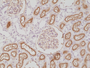
Immunohistochemical staining of formalin fixed and paraffin embedded human kidney tissue section using anti-CD10 rabbit monoclonal antibody (Clone RM337) at a 1:200 dilution.
Immunohistochemical staining of formalin fixed and paraffin embedded human kidney tissue section using anti-CD10 rabbit monoclonal antibody (Clone RM337) at a 1:200 dilution.
anti-CD10 (human), Rabbit Monoclonal (RM337)
REV-31-1224-00
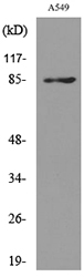
ApplicationsWestern Blot, ImmunoHistoChemistry
Product group Antibodies
ReactivityHuman
TargetMME
Overview
- SupplierRevMAb Biosciences
- Product Nameanti-CD10 (human), Rabbit Monoclonal (RM337)
- Delivery Days Customer2
- ApplicationsWestern Blot, ImmunoHistoChemistry
- CertificationResearch Use Only
- ClonalityMonoclonal
- Clone IDRM337
- Gene ID4311
- Target nameMME
- Target descriptionmembrane metalloendopeptidase
- Target synonymsCALLA, CD10, CMT2T, NEP, SCA43, SFE, neprilysin, atriopeptidase, common acute lymphocytic leukemia antigen, membrane metallo-endopeptidase (neutral endopeptidase, enkephalinase, CALLA, CD10), neprilysin-390, neprilysin-411, neutral endopeptidase 24.11, skin fibroblast elastase
- HostRabbit
- IsotypeIgG
- Protein IDP08473
- Protein NameNeprilysin
- Scientific DescriptionCD10 (Neprilysin; Common Acute Lymphocytic Leukemia Antigen; CALLA), is a cell surface enzyme with neutral metalloendopeptidase activity which inactivates a variety of biologically active peptides. CD10 is expressed in a wide variety of tissues and is particularly abundant in kidney. It is also a common acute lymphocytic leukemia antigen that is an important cell surface marker in the diagnosis of human acute lymphocytic leukemia (ALL) and several other lymphomas or epithelial tumors. CD10 is also present on breast myoepithelial cells, bile canaliculi, fibroblasts, with especially high expression on the brush border of kidney and gut epithelial cells. CD10 is a zinc-dependent metalloprotease that cleaves peptides at the amino side of hydrophobic residues and inactivates several peptide hormones including glucagon, enkephalins, substance P, neurotensin, oxytocin and bradykinin. Diseases associated with CD10 dysfunction include spinocerebellar ataxia 43 and Charcot-Marie tooth Disease. - Recombinant Antibody. This antibody reacts to the cytoplasmic domain of human CD10. Applications: WB, IHC. Source: Rabbit. Liquid. 50% Glycerol/PBS with 1% BSA and 0.09% sodium azide. CD10 (Neprilysin; Common Acute Lymphocytic Leukemia Antigen; CALLA), is a cell surface enzyme with neutral metalloendopeptidase activity which inactivates a variety of biologically active peptides. CD10 is expressed in a wide variety of tissues and is particularly abundant in kidney. It is also a common acute lymphocytic leukemia antigen that is an important cell surface marker in the diagnosis of human acute lymphocytic leukemia (ALL) and several other lymphomas or epithelial tumors. CD10 is also present on breast myoepithelial cells, bile canaliculi, fibroblasts, with especially high expression on the brush border of kidney and gut epithelial cells. CD10 is a zinc-dependent metalloprotease that cleaves peptides at the amino side of hydrophobic residues and inactivates several peptide hormones including glucagon, enkephalins, substance P, neurotensin, oxytocin and bradykinin. Diseases associated with CD10 dysfunction include spinocerebellar ataxia 43 and Charcot-Marie tooth Disease.
- ReactivityHuman
- Storage Instruction-20°C,2°C to 8°C
- UNSPSC41116161
Datasheet
Related products
Product group Antibodies
Anti-MME AntibodyA98473
ApplicationsWestern Blot, ELISA
ReactivityHuman, Mouse, Rat
- SizePrice

Product group Antibodies
Anti-MME AntibodyAMAB91788
ApplicationsImmunoHistoChemistry
ReactivityHuman
TargetMME
- SizePrice
Product group Antibodies

MME / CD10 Antibody (clone CB-CALLA)LS-C769989
ApplicationsFlow Cytometry
ReactivityHuman
TargetMME
- SizePrice
Product group Antibodies

Anti-CD10 [NL-1]Ab00950-10.0
ApplicationsImmunoFluorescence, ImmunoPrecipitation, Western Blot
ReactivityHuman
TargetMME
- SizePrice
Product group Antibodies
References
CD10 Polyclonal AntibodyBS-0527R
ApplicationsWestern Blot
ReactivityHuman, Mouse, Rat
TargetMME
- SizePrice
Product group Antibodies
MME Monoclonal AntibodyCSB-MA000230
ApplicationsELISA, ImmunoHistoChemistry
ReactivityHuman, Mouse, Rat
TargetMME
- SizePrice
Product group Antibodies
ApplicationsFlow Cytometry
TargetMME
- SizePrice
![CD10 antibody [N2C1], Internal detects MME protein by western blot analysis. A. 50 μg rat kidney lysate/extract 7.5% SDS-PAGE CD10 antibody [N2C1], Internal (GTX111680) dilution: 1:500 The HRP-conjugated anti-rabbit IgG antibody (GTX213110-01) was used to detect the primary antibody.](https://www.genetex.com/upload/website/prouct_img/normal/GTX111680/GTX111680_40485_WB_R_kidney_w_23060500_651.webp)
Product group Antibodies
CD10 antibody [N2C1], InternalGTX111680
ApplicationsWestern Blot
ReactivityHuman, Mouse, Rat
TargetMME
- SizePrice