

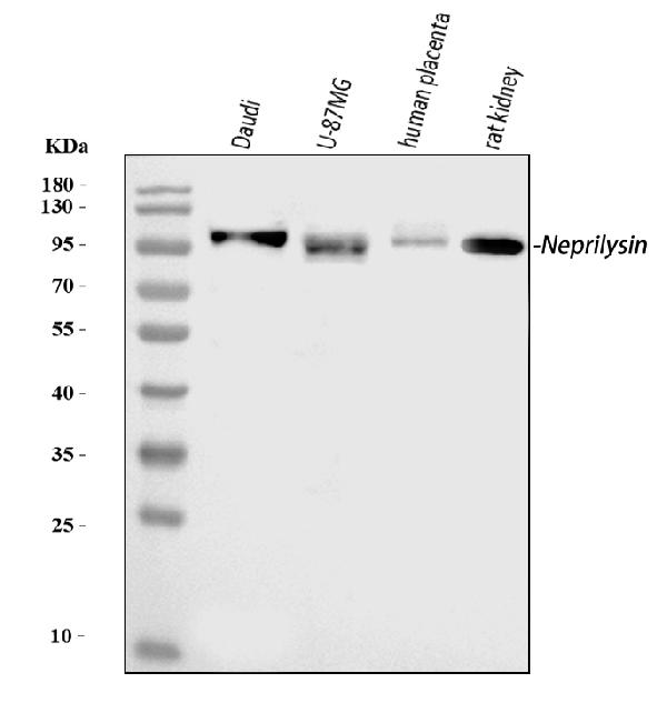
Figure 1. Western blot analysis of CD10/MME using anti-CD10/MME antibody (PB9058). Electrophoresis was performed on a 5-20% SDS-PAGE gel at 70V (Stacking gel) / 90V (Resolving gel) for 2-3 hours. The sample well of each lane was loaded with 30 ug of sample under reducing conditions. Lane 1: human Daudi whole cell lysates, Lane 2: human U-87MG whole cell lysates, Lane 3: human placenta tissue lysates, Lane 4: rat kidney tissue lysates. After electrophoresis, proteins were transferred to a nitrocellulose membrane at 150 mA for 50-90 minutes. Blocked the membrane with 5% non-fat milk/TBS for 1.5 hour at RT. The membrane was incubated with rabbit anti-CD10/MME antigen affinity purified polyclonal antibody (Catalog # PB9058) at 0.5 microg/mL overnight at 4°C, then washed with TBS-0.1%Tween 3 times with 5 minutes each and probed with a goat anti-rabbit IgG-HRP secondary antibody at a dilution of 1:5000 for 1.5 hour at RT. The signal is developed using an Enhanced Chemiluminescent detection (ECL) kit (Catalog # EK1002) with Tanon 5200 system. A specific band was detected for CD10/MME at approximately 100 kDa. The expected band size for CD10/MME is at 85 kDa.
. CD10/MME was detected in a paraffin-embedded section of human lymphoma tissue. Heat mediated antigen retrieval was performed in EDTA buffer (pH 8.0, epitope retrieval solution). The tissue section was blocked with 10% goat serum. The tissue section was then incubated with 2 microg/ml rabbit anti-CD10/MME Antibody (PB9058) overnight at 4°C. Peroxidase Conjugated Goat Anti-rabbit IgG was used as secondary antibody and incubated for 30 minutes at 37°C. The tissue section was developed using HRP Conjugated Rabbit IgG Super Vision Assay Kit (Catalog # SV0002) with DAB as the chromogen.")
. CD10/MME was detected in a paraffin-embedded section of human placenta tissue. Heat mediated antigen retrieval was performed in EDTA buffer (pH 8.0, epitope retrieval solution). The tissue section was blocked with 10% goat serum. The tissue section was then incubated with 2 microg/ml rabbit anti-CD10/MME Antibody (PB9058) overnight at 4°C. Peroxidase Conjugated Goat Anti-rabbit IgG was used as secondary antibody and incubated for 30 minutes at 37°C. The tissue section was developed using HRP Conjugated Rabbit IgG Super Vision Assay Kit (Catalog # SV0002) with DAB as the chromogen.")
. CD10/MME was detected in a paraffin-embedded section of human renal cell carcinoma tissue. Heat mediated antigen retrieval was performed in EDTA buffer (pH 8.0, epitope retrieval solution). The tissue section was blocked with 10% goat serum. The tissue section was then incubated with 2 microg/ml rabbit anti-CD10/MME Antibody (PB9058) overnight at 4°C. Peroxidase Conjugated Goat Anti-rabbit IgG was used as secondary antibody and incubated for 30 minutes at 37°C. The tissue section was developed using HRP Conjugated Rabbit IgG Super Vision Assay Kit (Catalog # SV0002) with DAB as the chromogen.")
. Overlay histogram showing Daudi cells stained with PB9058 (Blue line). The cells were fixed with 4% paraformaldehyde and blocked with 10% normal goat serum. And then incubated with rabbit anti-CD10/MME Antibody (PB9058, 1 microg/1x106 cells) for 30 min at 20°C. DyLight®488 conjugated goat anti-rabbit IgG (BA1127, 5-10 microg/1x106 cells) was used as secondary antibody for 30 minutes at 20°C. Isotype control antibody (Green line) was rabbit IgG (1 microg/1x106) used under the same conditions. Unlabelled sample without incubation with primary antibody and secondary antibody (Red line) was used as a blank control.")
Figure 1. Western blot analysis of CD10/MME using anti-CD10/MME antibody (PB9058). Electrophoresis was performed on a 5-20% SDS-PAGE gel at 70V (Stacking gel) / 90V (Resolving gel) for 2-3 hours. The sample well of each lane was loaded with 30 ug of sample under reducing conditions. Lane 1: human Daudi whole cell lysates, Lane 2: human U-87MG whole cell lysates, Lane 3: human placenta tissue lysates, Lane 4: rat kidney tissue lysates. After electrophoresis, proteins were transferred to a nitrocellulose membrane at 150 mA for 50-90 minutes. Blocked the membrane with 5% non-fat milk/TBS for 1.5 hour at RT. The membrane was incubated with rabbit anti-CD10/MME antigen affinity purified polyclonal antibody (Catalog # PB9058) at 0.5 microg/mL overnight at 4°C, then washed with TBS-0.1%Tween 3 times with 5 minutes each and probed with a goat anti-rabbit IgG-HRP secondary antibody at a dilution of 1:5000 for 1.5 hour at RT. The signal is developed using an Enhanced Chemiluminescent detection (ECL) kit (Catalog # EK1002) with Tanon 5200 system. A specific band was detected for CD10/MME at approximately 100 kDa. The expected band size for CD10/MME is at 85 kDa.
Anti-CD10/MME Antibody Picoband(r)
PB9058-CARRIER-FREE
ApplicationsFlow Cytometry, Western Blot, ImmunoHistoChemistry
Product group Antibodies
ReactivityHuman, Rat
TargetMME
Overview
- SupplierBoster Bio
- Product NameAnti-CD10/MME Antibody Picoband(r)
- Delivery Days Customer9
- Application Supplier NoteWB: The detection limit for CD10 is approximately 0.25ng/lane under reducing conditions. Tested Species: In-house tested species with positive results. By Heat: Boiling the paraffin sections in 10mM citrate buffer, pH6.0, for 20mins is required for the staining of formalin/paraffin sections. Other applications have not been tested. Optimal dilutions should be determined by end users.
- ApplicationsFlow Cytometry, Western Blot, ImmunoHistoChemistry
- CertificationResearch Use Only
- ClonalityPolyclonal
- Concentration500 ug/ml
- Gene ID4311
- Target nameMME
- Target descriptionmembrane metalloendopeptidase
- Target synonymsCALLA, CD10, CMT2T, NEP, SCA43, SFE, neprilysin, atriopeptidase, common acute lymphocytic leukemia antigen, membrane metallo-endopeptidase (neutral endopeptidase, enkephalinase, CALLA, CD10), neprilysin-390, neprilysin-411, neutral endopeptidase 24.11, skin fibroblast elastase
- HostRabbit
- IsotypeIgG
- Protein IDP08473
- Protein NameNeprilysin
- Scientific DescriptionBoster Bio Anti-CD10/MME Antibody Picoband® catalog # PB9058. Tested in Flow Cytometry, IHC, WB applications. This antibody reacts with Human, Rat. The brand Picoband indicates this is a premium antibody that guarantees superior quality, high affinity, and strong signals with minimal background in Western blot applications. Only our best-performing antibodies are designated as Picoband, ensuring unmatched performance.
- ReactivityHuman, Rat
- Storage Instruction-20°C,2°C to 8°C
- UNSPSC12352203
Related products
Product group Antibodies
ApplicationsFlow Cytometry
ReactivityHuman
TargetMME
- SizePrice

Product group Antibodies
Anti-MME AntibodyAMAB91788
ApplicationsImmunoHistoChemistry
ReactivityHuman
TargetMME
- SizePrice
Product group Antibodies

Anti-CD10 [NL-1]Ab00950-10.0
ApplicationsImmunoFluorescence, ImmunoPrecipitation, Western Blot
ReactivityHuman
TargetMME
- SizePrice
Product group Antibodies
Anti-MME AntibodyA98473
ApplicationsWestern Blot, ELISA
ReactivityHuman, Mouse, Rat
- SizePrice
Product group Antibodies

MME / CD10 Antibody (clone CB-CALLA)LS-C769989
ApplicationsFlow Cytometry
ReactivityHuman
TargetMME
- SizePrice
Product group Antibodies
MME Monoclonal AntibodyCSB-MA000230
ApplicationsELISA, ImmunoHistoChemistry
ReactivityHuman, Mouse, Rat
TargetMME
- SizePrice
Product group Antibodies
ApplicationsFlow Cytometry
TargetMME
- SizePrice
![CD10 antibody [N2C1], Internal detects MME protein by western blot analysis. A. 50 μg rat kidney lysate/extract 7.5% SDS-PAGE CD10 antibody [N2C1], Internal (GTX111680) dilution: 1:500 The HRP-conjugated anti-rabbit IgG antibody (GTX213110-01) was used to detect the primary antibody.](https://www.genetex.com/upload/website/prouct_img/normal/GTX111680/GTX111680_40485_WB_R_kidney_w_23060500_651.webp)
Product group Antibodies
CD10 antibody [N2C1], InternalGTX111680
ApplicationsWestern Blot
ReactivityHuman, Mouse, Rat
TargetMME
- SizePrice
Product group Antibodies
References
CD10 Polyclonal AntibodyBS-0527R
ApplicationsWestern Blot
ReactivityHuman, Mouse, Rat
TargetMME
- SizePrice