

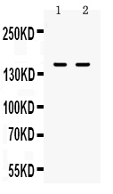
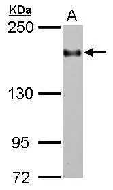
Figure 1. Western blot analysis of CD11a using anti-CD11a antibody (A04466-1). Electrophoresis was performed on a 5-20% SDS-PAGE gel at 70V (Stacking gel) / 90V (Resolving gel) for 2-3 hours. The sample well of each lane was loaded with 50ug of sample under reducing conditions. lane 1: JURKAT cell lysates, lane 2: CEM whole cell lysates. After Electrophoresis, proteins were transferred to a Nitrocellulose membrane at 150mA for 50-90 minutes. Blocked the membrane with 5% Non-fat Milk/ TBS for 1.5 hour at RT. The membrane was incubated with rabbit anti-CD11a antigen affinity purified polyclonal antibody (Catalog # A04466-1) at 0.5 microg/mL overnight at 4°C, then washed with TBS-0.1%Tween 3 times with 5 minutes each and probed with a goat anti-rabbit IgG-HRP secondary antibody at a dilution of 1:10000 for 1.5 hour at RT. The signal is developed using an Enhanced Chemiluminescent detection (ECL) kit (Catalog # EK1002) with Tanon 5200 system. A specific band was detected for CD11a at approximately 150KD. The expected band size for CD11a is at 129KD.
. Overlay histogram showing U937 cells stained with A04466-1 (Blue line).The cells were blocked with 10% normal goat serum. And then incubated with rabbit anti-CD11a Antibody (A04466-1,1microg/1x106 cells) for 30 min at 20°C. DyLight488 conjugated goat anti-rabbit IgG (BA1127, 5-10microg/1x106 cells) was used as secondary antibody for 30 minutes at 20°C. Isotype control antibody (Green line) was rabbit IgG (1microg/1x106) used under the same conditions. Unlabelled sample (Red line) was also used as a control.")
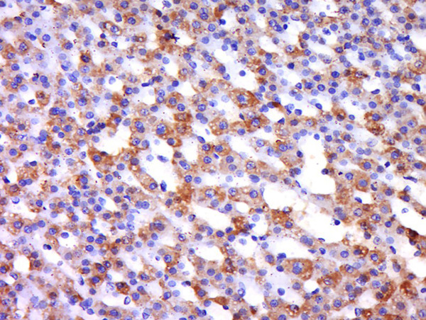
. CD11a was detected in immunocytochemical section of U937 Cell. Enzyme antigen retrieval was performed using IHC enzyme antigen retrieval reagent (AR0022) for 15 mins. The cells were blocked with 10% goat serum. And then incubated with 1microg/ml rabbit anti-CD11a Antibody (A04466-1) overnight at 4°C. Biotinylated goat anti-rabbit IgG was used as secondary antibody and incubated for 30 minutes at 37°C. The section was developed using Strepavidin-Biotin-Complex (SABC)(Catalog # SA1022) with DAB as the chromogen.")
Figure 1. Western blot analysis of CD11a using anti-CD11a antibody (A04466-1). Electrophoresis was performed on a 5-20% SDS-PAGE gel at 70V (Stacking gel) / 90V (Resolving gel) for 2-3 hours. The sample well of each lane was loaded with 50ug of sample under reducing conditions. lane 1: JURKAT cell lysates, lane 2: CEM whole cell lysates. After Electrophoresis, proteins were transferred to a Nitrocellulose membrane at 150mA for 50-90 minutes. Blocked the membrane with 5% Non-fat Milk/ TBS for 1.5 hour at RT. The membrane was incubated with rabbit anti-CD11a antigen affinity purified polyclonal antibody (Catalog # A04466-1) at 0.5 microg/mL overnight at 4°C, then washed with TBS-0.1%Tween 3 times with 5 minutes each and probed with a goat anti-rabbit IgG-HRP secondary antibody at a dilution of 1:10000 for 1.5 hour at RT. The signal is developed using an Enhanced Chemiluminescent detection (ECL) kit (Catalog # EK1002) with Tanon 5200 system. A specific band was detected for CD11a at approximately 150KD. The expected band size for CD11a is at 129KD.
Anti-CD11a/ITGAL Antibody Picoband(r)
A04466-1-CARRIER-FREE
ApplicationsFlow Cytometry, Western Blot, ImmunoCytoChemistry, ImmunoHistoChemistry
Product group Antibodies
ReactivityHuman
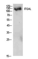
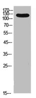
TargetITGAL
Overview
- SupplierBoster Bio
- Product NameAnti-CD11a/ITGAL Antibody Picoband(r)
- Delivery Days Customer9
- ApplicationsFlow Cytometry, Western Blot, ImmunoCytoChemistry, ImmunoHistoChemistry
- CertificationResearch Use Only
- ClonalityPolyclonal
- Concentration500 ug/ml
- Gene ID3683
- Target nameITGAL
- Target descriptionintegrin subunit alpha L
- Target synonymsCD11A, LFA-1, LFA1A, integrin alpha-L, CD11 antigen-like family member A, LFA-1 alpha, LFA-1A, antigen CD11A (p180), lymphocyte function-associated antigen 1, alpha polypeptide, integrin gene promoter, integrin, alpha L (antigen CD11A (p180), lymphocyte function-associated antigen 1; alpha polypeptide), leukocyte adhesion glycoprotein LFA-1 alpha chain, leukocyte function-associated molecule 1 alpha chain, lymphocyte function-associated antigen 1, alpha polypeptide
- HostRabbit
- IsotypeIgG
- Protein IDP20701
- Protein NameIntegrin alpha-L
- Scientific DescriptionBoster Bio Anti-CD11a/ITGAL Antibody Picoband® catalog # A04466-1. Tested in Flow Cytometry, IHC, ICC, WB applications. This antibody reacts with Human. The brand Picoband indicates this is a premium antibody that guarantees superior quality, high affinity, and strong signals with minimal background in Western blot applications. Only our best-performing antibodies are designated as Picoband, ensuring unmatched performance.
- ReactivityHuman
- Storage Instruction-20°C,2°C to 8°C
- UNSPSC12352203
Related products
Product group Antibodies

Anti-CD11 alpha/Integrin alpha-L [YTH 81.5]Ab00190-1.1
ApplicationsFlow Cytometry
ReactivityHuman
TargetITGAL
- SizePrice
Product group Antibodies
Anti-ITGAL AntibodyA101010
ApplicationsWestern Blot, ELISA
ReactivityHuman
- SizePrice
Product group Antibodies
Anti-ITGAL Antibody144-01644
ApplicationsWestern Blot, ImmunoHistoChemistry
ReactivityHuman, Mouse, Rat
TargetITGAL
- SizePrice
Product group Antibodies

ITGAL / CD11a Antibody (clone FD441.8)LS-C812186
ApplicationsFlow Cytometry
ReactivityMouse
TargetITGAL
- SizePrice
Product group Antibodies
References
CD11a Polyclonal AntibodyBS-1804R
ApplicationsFlow Cytometry, ImmunoFluorescence, ImmunoCytoChemistry, ImmunoHistoChemistry, ImmunoHistoChemistry Frozen, ImmunoHistoChemistry Paraffin
ReactivityBovine, Canine, Equine, Human, Mouse, Porcine, Rabbit, Rat, Sheep
TargetITGAL
- SizePrice
Product group Antibodies
ITGAL AntibodyCSB-PA006157
ApplicationsWestern Blot, ELISA
ReactivityHuman
TargetITGAL
- SizePrice
Product group Antibodies

ApplicationsWestern Blot, ELISA, ImmunoHistoChemistry
ReactivityHuman
TargetITGAL
- SizePrice
Product group Antibodies
Itgal Polyclonal AntibodyCAC08566
ApplicationsImmunoFluorescence, Western Blot, ELISA, ImmunoHistoChemistry
TargetITGAL
- SizePrice
Product group Antibodies
CD11a antibody [C1C2], InternalGTX101564
ApplicationsWestern Blot
ReactivityHuman
TargetITGAL
- SizePrice