

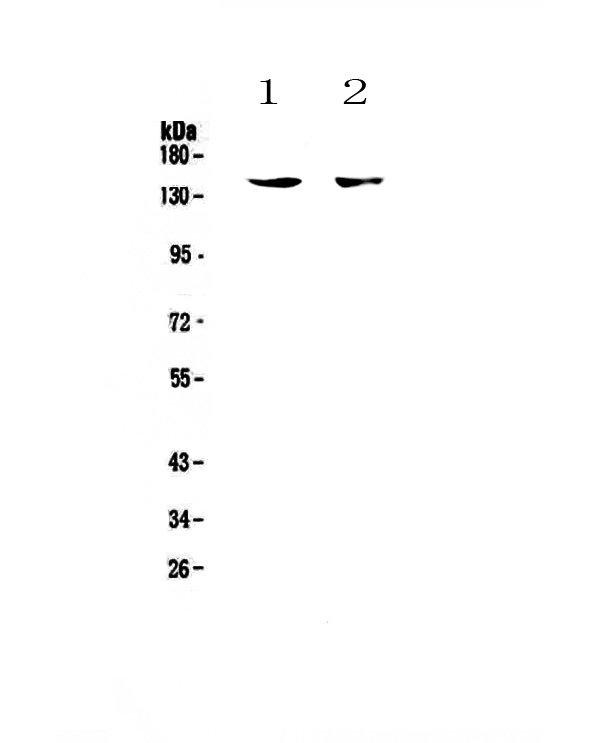
Figure 1. Western blot analysis of CD11c using anti-CD11c antibody (A00357-1). Electrophoresis was performed on a 5-20% SDS-PAGE gel at 70V (Stacking gel) / 90V (Resolving gel) for 2-3 hours. The sample well of each lane was loaded with 50ug of sample under reducing conditions. Lane 1: mouse spleen tissue lysate, Lane 2: mouse thymus tissue lysate. After Electrophoresis, proteins were transferred to a Nitrocellulose membrane at 150mA for 50-90 minutes. Blocked the membrane with 5% Non-fat Milk/ TBS for 1.5 hour at RT. The membrane was incubated with rabbit anti-CD11c antigen affinity purified polyclonal antibody (Catalog # A00357-1) at 0.5 microg/mL overnight at 4°C, then washed with TBS-0.1%Tween 3 times with 5 minutes each and probed with a goat anti-rabbit IgG-HRP secondary antibody at a dilution of 1:10000 for 1.5 hour at RT. The signal is developed using an Enhanced Chemiluminescent detection (ECL) kit (Catalog # EK1002) with Tanon 5200 system. A specific band was detected for CD11c at approximately 150KD. The expected band size for CD11c is at 128KD.
. CD11c was detected in paraffin-embedded section of human tonsil tissue . Heat mediated antigen retrieval was performed in citrate buffer (pH6, epitope retrieval solution) for 20 mins. The tissue section was blocked with 10% goat serum. The tissue section was then incubated with 1microg/ml rabbit anti-CD11c Antibody (A00357-1) overnight at 4°C. Biotinylated goat anti-rabbit IgG was used as secondary antibody and incubated for 30 minutes at 37°C. The tissue section was developed using Strepavidin-Biotin-Complex (SABC)(Catalog # SA1022) with DAB as the chromogen.")
Figure 1. Western blot analysis of CD11c using anti-CD11c antibody (A00357-1). Electrophoresis was performed on a 5-20% SDS-PAGE gel at 70V (Stacking gel) / 90V (Resolving gel) for 2-3 hours. The sample well of each lane was loaded with 50ug of sample under reducing conditions. Lane 1: mouse spleen tissue lysate, Lane 2: mouse thymus tissue lysate. After Electrophoresis, proteins were transferred to a Nitrocellulose membrane at 150mA for 50-90 minutes. Blocked the membrane with 5% Non-fat Milk/ TBS for 1.5 hour at RT. The membrane was incubated with rabbit anti-CD11c antigen affinity purified polyclonal antibody (Catalog # A00357-1) at 0.5 microg/mL overnight at 4°C, then washed with TBS-0.1%Tween 3 times with 5 minutes each and probed with a goat anti-rabbit IgG-HRP secondary antibody at a dilution of 1:10000 for 1.5 hour at RT. The signal is developed using an Enhanced Chemiluminescent detection (ECL) kit (Catalog # EK1002) with Tanon 5200 system. A specific band was detected for CD11c at approximately 150KD. The expected band size for CD11c is at 128KD.
Anti-CD11c/ITGAX Antibody Picoband(r)
A00357-1-CARRIER-FREE
ApplicationsWestern Blot, ELISA, ImmunoHistoChemistry
Product group Antibodies
ReactivityHuman, Mouse
TargetITGAX
Overview
- SupplierBoster Bio
- Product NameAnti-CD11c/ITGAX Antibody Picoband(r)
- Delivery Days Customer9
- ApplicationsWestern Blot, ELISA, ImmunoHistoChemistry
- CertificationResearch Use Only
- ClonalityPolyclonal
- Concentration500 ug/ml
- Gene ID3687
- Target nameITGAX
- Target descriptionintegrin subunit alpha X
- Target synonymsCD11C, SLEB6, integrin alpha-X, CD11 antigen-like family member C, complement component 3 receptor 4 subunit, integrin alpha X, integrin, alpha X (antigen CD11C (p150), alpha polypeptide), integrin, alpha X (complement component 3 receptor 4 subunit), leu M5, alpha subunit, leukocyte adhesion glycoprotein p150,95 alpha chain, leukocyte adhesion receptor p150,95, leukocyte surface antigen p150,95, alpha subunit, myeloid membrane antigen, alpha subunit, p150 95 integrin alpha chain
- HostRabbit
- IsotypeIgG
- Protein IDP20702
- Protein NameIntegrin alpha-X
- Scientific DescriptionBoster Bio Anti-CD11c/ITGAX Antibody Picoband® catalog # A00357-1. Tested in ELISA, IHC, WB applications. This antibody reacts with Human, Mouse. The brand Picoband indicates this is a premium antibody that guarantees superior quality, high affinity, and strong signals with minimal background in Western blot applications. Only our best-performing antibodies are designated as Picoband, ensuring unmatched performance.
- ReactivityHuman, Mouse
- Storage Instruction-20°C,2°C to 8°C
- UNSPSC12352203
Related products
Product group Antibodies
ITGAX AntibodyCSB-PA011879ESR1HU
ApplicationsImmunoFluorescence, Western Blot, ELISA, ImmunoHistoChemistry
ReactivityHuman, Mouse
TargetITGAX
- SizePrice
Product group Antibodies
Anti-Human CD11 C Antibody, PE136-18137
ApplicationsFlow Cytometry
ReactivityHuman
TargetITGAX
- SizePrice


Product group Antibodies
Anti-ITGAX AntibodyAMAB90915
ApplicationsWestern Blot, ImmunoHistoChemistry
ReactivityHuman
TargetITGAX
- SizePrice
Product group Antibodies

Anti-CD11c [N418]Ab00420-1.1
ApplicationsFlow Cytometry, ImmunoFluorescence, Other Application
ReactivityHuman, Mouse
TargetITGAX
- SizePrice
Product group Antibodies

ITGAX / CD11c Antibody (clone N418, PE)LS-C764332
ApplicationsFlow Cytometry
ReactivityMouse
TargetITGAX
- SizePrice
Product group Antibodies
Itgax Polyclonal AntibodyCAC10873
ApplicationsImmunoFluorescence, Western Blot, ELISA, ImmunoHistoChemistry
ReactivityMouse
TargetITGAX
- SizePrice