


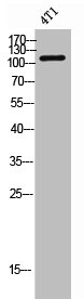
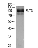
Figure 1. Western blot analysis of CD135/FLT3 using anti-CD135/FLT3 antibody (A00188-5). Electrophoresis was performed on a 5-20% SDS-PAGE gel at 70V (Stacking gel) / 90V (Resolving gel) for 2-3 hours. The sample well of each lane was loaded with 30 ug of sample under reducing conditions. Lane 1: human HCCP tissue lysates. After electrophoresis, proteins were transferred to a nitrocellulose membrane at 150 mA for 50-90 minutes. Blocked the membrane with 5% non-fat milk/TBS for 1.5 hour at RT. The membrane was incubated with rabbit anti-CD135/FLT3 antigen affinity purified polyclonal antibody (Catalog # A00188-5) at 0.5 microg/mL overnight at 4°C, then washed with TBS-0.1%Tween 3 times with 5 minutes each and probed with a goat anti-rabbit IgG-HRP secondary antibody at a dilution of 1:5000 for 1.5 hour at RT. The signal is developed using an Enhanced Chemiluminescent detection (ECL) kit (Catalog # EK1002) with Tanon 5200 system. A specific band was detected for CD135/FLT3 at approximately 160 kDa. The expected band size for CD135/FLT3 is at 113 kDa.
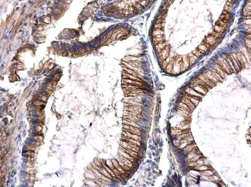
. CD135/FLT3 was detected in a paraffin-embedded section of human spleen tissue. Heat mediated antigen retrieval was performed in EDTA buffer (pH 8.0, epitope retrieval solution). The tissue section was blocked with 10% goat serum. The tissue section was then incubated with 2 microg/ml rabbit anti-CD135/FLT3 Antibody (A00188-5) overnight at 4°C. Peroxidase Conjugated Goat Anti-rabbit IgG was used as secondary antibody and incubated for 30 minutes at 37°C. The tissue section was developed using HRP Conjugated Rabbit IgG Super Vision Assay Kit (Catalog # SV0002) with DAB as the chromogen.")
Figure 1. Western blot analysis of CD135/FLT3 using anti-CD135/FLT3 antibody (A00188-5). Electrophoresis was performed on a 5-20% SDS-PAGE gel at 70V (Stacking gel) / 90V (Resolving gel) for 2-3 hours. The sample well of each lane was loaded with 30 ug of sample under reducing conditions. Lane 1: human HCCP tissue lysates. After electrophoresis, proteins were transferred to a nitrocellulose membrane at 150 mA for 50-90 minutes. Blocked the membrane with 5% non-fat milk/TBS for 1.5 hour at RT. The membrane was incubated with rabbit anti-CD135/FLT3 antigen affinity purified polyclonal antibody (Catalog # A00188-5) at 0.5 microg/mL overnight at 4°C, then washed with TBS-0.1%Tween 3 times with 5 minutes each and probed with a goat anti-rabbit IgG-HRP secondary antibody at a dilution of 1:5000 for 1.5 hour at RT. The signal is developed using an Enhanced Chemiluminescent detection (ECL) kit (Catalog # EK1002) with Tanon 5200 system. A specific band was detected for CD135/FLT3 at approximately 160 kDa. The expected band size for CD135/FLT3 is at 113 kDa.
Anti-CD135/FLT3 Antibody Picoband(r)
A00188-5-CARRIER-FREE
ApplicationsWestern Blot, ELISA, ImmunoHistoChemistry
Product group Antibodies
ReactivityHuman
TargetFLT3
Overview
- SupplierBoster Bio
- Product NameAnti-CD135/FLT3 Antibody Picoband(r)
- Delivery Days Customer9
- ApplicationsWestern Blot, ELISA, ImmunoHistoChemistry
- CertificationResearch Use Only
- ClonalityPolyclonal
- Concentration500 ug/ml
- Gene ID2322
- Target nameFLT3
- Target descriptionfms related receptor tyrosine kinase 3
- Target synonymsCD135, FLK-2, FLK2, STK1, receptor-type tyrosine-protein kinase FLT3, CD135 antigen, FL cytokine receptor, fetal liver kinase 2, fms related tyrosine kinase 3, fms-like tyrosine kinase 3, growth factor receptor tyrosine kinase type III, stem cell tyrosine kinase 1
- HostRabbit
- IsotypeIgG
- Protein IDP36888
- Protein NameReceptor-type tyrosine-protein kinase FLT3
- Scientific DescriptionBoster Bio Anti-CD135/FLT3 Antibody Picoband® catalog # A00188-5. Tested in ELISA, IHC, WB applications. This antibody reacts with Human. The brand Picoband indicates this is a premium antibody that guarantees superior quality, high affinity, and strong signals with minimal background in Western blot applications. Only our best-performing antibodies are designated as Picoband, ensuring unmatched performance.
- ReactivityHuman
- Storage Instruction-20°C,2°C to 8°C
- UNSPSC12352203
Related products
Product group Antibodies
FLT3 AntibodyCSB-PA006265
ApplicationsWestern Blot, ELISA
ReactivityHuman, Mouse, Rat
TargetFLT3
- SizePrice
Product group Antibodies

Anti-FLT3 [EB10]Ab03003-10.0
ApplicationsFlow Cytometry, ELISA, Other Application
ReactivityHuman
TargetFLT3
- SizePrice
Product group Antibodies
Anti-FLT3 AntibodyA98148
ApplicationsWestern Blot, ELISA
ReactivityHuman, Mouse, Rat
- SizePrice
Product group Antibodies

CD135 / FLT3 AntibodyLS-C831609
ApplicationsWestern Blot, ImmunoHistoChemistry
ReactivityHuman, Mouse, Rat
TargetFLT3
- SizePrice

Product group Antibodies
Anti-FLT3 AntibodyHPA047539
ApplicationsImmunoCytoChemistry
ReactivityHuman
TargetFLT3
- SizePrice
Product group Antibodies
FLT3 Polyclonal AntibodyCAC15776
ApplicationsWestern Blot, ELISA
TargetFLT3
- SizePrice
Product group Antibodies
FLT3 Polyclonal AntibodyBS-2767R
ApplicationsImmunoFluorescence, Western Blot, ELISA, ImmunoCytoChemistry, ImmunoHistoChemistry, ImmunoHistoChemistry Frozen, ImmunoHistoChemistry Paraffin
ReactivityHuman, Mouse, Rat
TargetFLT3
- SizePrice
Product group Antibodies
FLT3 antibodyGTX101556
ApplicationsImmunoHistoChemistry, ImmunoHistoChemistry Paraffin
ReactivityHuman
TargetFLT3
- SizePrice