


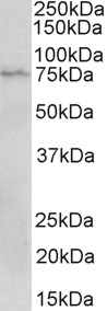
Figure 1. Western blot analysis of CD19 using anti-CD19 antibody (PB9800). Electrophoresis was performed on a 5-20% SDS-PAGE gel at 70V (Stacking gel) / 90V (Resolving gel) for 2-3 hours. The sample well of each lane was loaded with 30 ug of sample under reducing conditions. Lane 1: human Raji whole cell lysates, Lane 2: human Ramos whole cell lysates, Lane 3: human Daudi whole cell lysates. After electrophoresis, proteins were transferred to a nitrocellulose membrane at 150 mA for 50-90 minutes. Blocked the membrane with 5% non-fat milk/TBS for 1.5 hour at RT. The membrane was incubated with rabbit anti-CD19 antigen affinity purified polyclonal antibody (Catalog # PB9800) at 0.5 microg/mL overnight at 4°C, then washed with TBS-0.1%Tween 3 times with 5 minutes each and probed with a goat anti-rabbit IgG-HRP secondary antibody at a dilution of 1:5000 for 1.5 hour at RT. The signal is developed using an Enhanced Chemiluminescent detection (ECL) kit (Catalog # EK1002) with Tanon 5200 system. A specific band was detected for CD19 at approximately 95 kDa. The expected band size for CD19 is at 61 kDa.
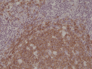
. CD19 was detected in a paraffin-embedded section of human appendix tissue. Heat mediated antigen retrieval was performed in EDTA buffer (pH 8.0, epitope retrieval solution). The tissue section was blocked with 10% goat serum. The tissue section was then incubated with 2 microg/ml rabbit anti-CD19 Antibody (PB9800) overnight at 4°C. Peroxidase Conjugated Goat Anti-rabbit IgG was used as secondary antibody and incubated for 30 minutes at 37°C. The tissue section was developed using HRP Conjugated Rabbit IgG Super Vision Assay Kit (Catalog # SV0002) with DAB as the chromogen.")
Figure 1. Western blot analysis of CD19 using anti-CD19 antibody (PB9800). Electrophoresis was performed on a 5-20% SDS-PAGE gel at 70V (Stacking gel) / 90V (Resolving gel) for 2-3 hours. The sample well of each lane was loaded with 30 ug of sample under reducing conditions. Lane 1: human Raji whole cell lysates, Lane 2: human Ramos whole cell lysates, Lane 3: human Daudi whole cell lysates. After electrophoresis, proteins were transferred to a nitrocellulose membrane at 150 mA for 50-90 minutes. Blocked the membrane with 5% non-fat milk/TBS for 1.5 hour at RT. The membrane was incubated with rabbit anti-CD19 antigen affinity purified polyclonal antibody (Catalog # PB9800) at 0.5 microg/mL overnight at 4°C, then washed with TBS-0.1%Tween 3 times with 5 minutes each and probed with a goat anti-rabbit IgG-HRP secondary antibody at a dilution of 1:5000 for 1.5 hour at RT. The signal is developed using an Enhanced Chemiluminescent detection (ECL) kit (Catalog # EK1002) with Tanon 5200 system. A specific band was detected for CD19 at approximately 95 kDa. The expected band size for CD19 is at 61 kDa.
Anti-CD19 Antibody Picoband(r)
PB9800
ApplicationsWestern Blot, ImmunoHistoChemistry
Product group Antibodies
ReactivityHuman
TargetCD19
Overview
- SupplierBoster Bio
- Product NameAnti-CD19 Antibody Picoband(r)
- Delivery Days Customer9
- Application Supplier NoteTested Species: In-house tested species with positive results. By Heat: Boiling the paraffin sections in 10mM citrate buffer, pH6.0, for 20mins is required for the staining of formalin/paraffin sections. Other applications have not been tested. Optimal dilutions should be determined by end users.
- ApplicationsWestern Blot, ImmunoHistoChemistry
- CertificationResearch Use Only
- ClonalityPolyclonal
- Concentration500 ug/ml
- Gene ID930
- Target nameCD19
- Target descriptionCD19 molecule
- Target synonymsB4, CVID3, B-lymphocyte antigen CD19, B-lymphocyte surface antigen B4, T-cell surface antigen Leu-12, differentiation antigen CD19
- HostRabbit
- IsotypeIgG
- Protein IDP15391
- Protein NameB-lymphocyte antigen CD19
- Scientific DescriptionBoster Bio Anti-CD19 Antibody Picoband® catalog # PB9800. Tested in IHC, WB applications. This antibody reacts with Human. The brand Picoband indicates this is a premium antibody that guarantees superior quality, high affinity, and strong signals with minimal background in Western blot applications. Only our best-performing antibodies are designated as Picoband, ensuring unmatched performance.
- ReactivityHuman
- Storage Instruction-20°C,2°C to 8°C
- UNSPSC12352203
Datasheet
MSDS
Related products
Product group Antibodies

Anti-CD19 [6D5]AB01489-2.0-VXS
ApplicationsFlow Cytometry, ImmunoPrecipitation, Western Blot, ImmunoHistoChemistry
ReactivityMouse
TargetCD19
- SizePrice
Product group Antibodies
ApplicationsFlow Cytometry
TargetCD19
- SizePrice
Product group Antibodies
Anti-CD19 Antibody144-02577
ApplicationsWestern Blot, ImmunoHistoChemistry
ReactivityHuman, Mouse, Rat
TargetCD19
- SizePrice

Product group Antibodies
Anti-CD19 Antibody Picoband(r)A00154-1-CARRIER-FREE
ApplicationsWestern Blot, ELISA
ReactivityHuman, Mouse
TargetCD19
- SizePrice
![FACS analysis of human peripheral blood lymphocytes using GTX01455-08 CD19 antibody [HIB19] (PE). Solid lone : primary antibody Dashed line : isotype control antibody amount : 0.25 μg (5 μl)](https://www.genetex.com/upload/website/prouct_img/normal/GTX01455-08/GTX01455-08_20200428_FACS100_w_23053121_847.webp)
Product group Antibodies
References
CD19 antibody [HIB19] (PE)GTX01455-08
ApplicationsFlow Cytometry
ReactivityHuman
TargetCD19
- SizePrice
Product group Antibodies
ApplicationsImmunoPrecipitation, Western Blot, ImmunoCytoChemistry, ImmunoHistoChemistry
ReactivityMouse, Rat
TargetCD19
- SizePrice
Product group Antibodies
Anti-CD19 AntibodyA82935
ApplicationsWestern Blot, ELISA
ReactivityHuman
- SizePrice
Product group Antibodies
anti-CD19 (human), Rabbit Monoclonal (RM332)REV-31-1219-00
ApplicationsWestern Blot, ImmunoHistoChemistry
ReactivityHuman
TargetCD19
- SizePrice