


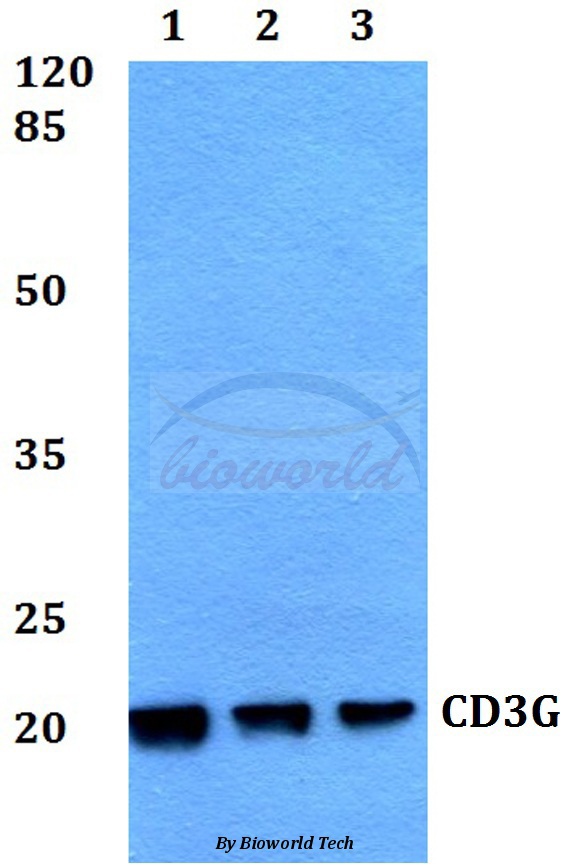
Figure 1. Western blot analysis of CD3 Gamma/CD3G using anti-CD3 Gamma/CD3G antibody (A04853-2). Electrophoresis was performed on a 5-20% SDS-PAGE gel at 70V (Stacking gel) / 90V (Resolving gel) for 2-3 hours. The sample well of each lane was loaded with 30 ug of sample under reducing conditions. Lane 1: human MOMLT-4 whole cell lysates, Lane 2: human Jurkat whole cell lysates. After electrophoresis, proteins were transferred to a nitrocellulose membrane at 150 mA for 50-90 minutes. Blocked the membrane with 5% non-fat milk/TBS for 1.5 hour at RT. The membrane was incubated with rabbit anti-CD3 Gamma/CD3G antigen affinity purified polyclonal antibody (Catalog # A04853-2) at 0.5 microg/mL overnight at 4°C, then washed with TBS-0.1%Tween 3 times with 5 minutes each and probed with a goat anti-rabbit IgG-HRP secondary antibody at a dilution of 1:5000 for 1.5 hour at RT. The signal is developed using an Enhanced Chemiluminescent detection (ECL) kit (Catalog # EK1002) with Tanon 5200 system. A specific band was detected for CD3 Gamma/CD3G at approximately 20-25 kDa. The expected band size for CD3 Gamma/CD3G is at 20-25 kDa.
. CD3 Gamma/CD3G was detected in an immunocytochemical section of JK cells. Enzyme antigen retrieval was performed using IHC enzyme antigen retrieval reagent (AR0022) for 15 mins. The cells were blocked with 10% goat serum. And then incubated with 5 microg/mL rabbit anti-CD3 Gamma/CD3G Antibody (A04853-2) overnight at 4°C. Cy3 Conjugated Goat Anti-Rabbit IgG (BA1032) was used as secondary antibody at 1:500 dilution and incubated for 30 minutes at 37°C. The section was counterstained with DAPI. Visualize using a fluorescence microscope and filter sets appropriate for the label used.")
. Overlay histogram showing JK cells stained with A04853-2 (Blue line). To facilitate intracellular staining, cells were fixed with 4% paraformaldehyde and permeabilized with permeabilization buffer. The cells were blocked with 10% normal goat serum. And then incubated with rabbit anti-CD3 Gamma/CD3G Antibody (A04853-2, 1 microg/1x106 cells) for 30 min at 20°C. DyLight®488 conjugated goat anti-rabbit IgG (BA1127, 5-10 microg/1x106 cells) was used as secondary antibody for 30 minutes at 20°C. Isotype control antibody (Green line) was rabbit IgG (1 microg/1x106) used under the same conditions. Unlabelled sample (Red line) was also used as a control.")
Figure 1. Western blot analysis of CD3 Gamma/CD3G using anti-CD3 Gamma/CD3G antibody (A04853-2). Electrophoresis was performed on a 5-20% SDS-PAGE gel at 70V (Stacking gel) / 90V (Resolving gel) for 2-3 hours. The sample well of each lane was loaded with 30 ug of sample under reducing conditions. Lane 1: human MOMLT-4 whole cell lysates, Lane 2: human Jurkat whole cell lysates. After electrophoresis, proteins were transferred to a nitrocellulose membrane at 150 mA for 50-90 minutes. Blocked the membrane with 5% non-fat milk/TBS for 1.5 hour at RT. The membrane was incubated with rabbit anti-CD3 Gamma/CD3G antigen affinity purified polyclonal antibody (Catalog # A04853-2) at 0.5 microg/mL overnight at 4°C, then washed with TBS-0.1%Tween 3 times with 5 minutes each and probed with a goat anti-rabbit IgG-HRP secondary antibody at a dilution of 1:5000 for 1.5 hour at RT. The signal is developed using an Enhanced Chemiluminescent detection (ECL) kit (Catalog # EK1002) with Tanon 5200 system. A specific band was detected for CD3 Gamma/CD3G at approximately 20-25 kDa. The expected band size for CD3 Gamma/CD3G is at 20-25 kDa.
Anti-CD3 Gamma/CD3G Antibody Picoband(r)
A04853-2-CARRIER-FREE
ApplicationsFlow Cytometry, ImmunoFluorescence, Western Blot, ELISA, ImmunoCytoChemistry
Product group Antibodies
ReactivityHuman
TargetCD3G
Overview
- SupplierBoster Bio
- Product NameAnti-CD3 Gamma/CD3G Antibody Picoband(r)
- Delivery Days Customer9
- ApplicationsFlow Cytometry, ImmunoFluorescence, Western Blot, ELISA, ImmunoCytoChemistry
- CertificationResearch Use Only
- ClonalityPolyclonal
- Concentration500 ug/ml
- Gene ID917
- Target nameCD3G
- Target descriptionCD3 gamma subunit of T-cell receptor complex
- Target synonymsCD3-GAMMA, CD3GAMMA, IMD17, T3G, T-cell surface glycoprotein CD3 gamma chain, CD3g antigen, gamma polypeptide (TiT3 complex), CD3g molecule, epsilon (CD3-TCR complex), CD3g molecule, gamma (CD3-TCR complex), T-cell antigen receptor complex, gamma subunit of T3, T-cell receptor T3 gamma chain
- HostRabbit
- IsotypeIgG
- Protein IDP09693
- Protein NameT-cell surface glycoprotein CD3 gamma chain
- Scientific DescriptionBoster Bio Anti-CD3 Gamma/CD3G Antibody Picoband® catalog # A04853-2. Tested in ELISA, Flow Cytometry, IF, ICC, WB applications. This antibody reacts with Human. The brand Picoband indicates this is a premium antibody that guarantees superior quality, high affinity, and strong signals with minimal background in Western blot applications. Only our best-performing antibodies are designated as Picoband, ensuring unmatched performance.
- ReactivityHuman
- Storage Instruction-20°C,2°C to 8°C
- UNSPSC12352203
Related products
Product group Antibodies
Anti-CD3G AntibodyA28542
ApplicationsWestern Blot
ReactivityHuman, Mouse, Rat
- SizePrice

Product group Antibodies
CD3 gamma Recombinant AntibodyBSM-54300R
ApplicationsFlow Cytometry, ImmunoFluorescence, Western Blot, ImmunoHistoChemistry, ImmunoHistoChemistry Frozen, ImmunoHistoChemistry Paraffin
ReactivityHuman, Mouse
TargetCD3G
- SizePrice
Product group Antibodies

Goat anti-CD3GEB12594
ApplicationsWestern Blot, ELISA
ReactivityBovine, Canine, Human, Mouse, Porcine, Rat
TargetCD3G
- SizePrice
Product group Antibodies
CD3G Polyclonal AntibodyCAC15040
ApplicationsImmunoFluorescence, Western Blot, ELISA
ReactivityMouse, Rat
TargetCD3G
- SizePrice
Product group Antibodies
CD3G AntibodyCSB-PA004933LA01HU
ApplicationsImmunoFluorescence, Western Blot, ELISA
ReactivityHuman, Mouse, Rat
TargetCD3G
- SizePrice
Product group Antibodies

CD3G AntibodyLS-C408379
ApplicationsFlow Cytometry, Western Blot, ImmunoHistoChemistry
ReactivityHuman
TargetCD3G
- SizePrice
![CD3 gamma antibody [C1C3] detects CD3 gamma protein at cell membrane by immunohistochemical analysis. Sample: Paraffin-embedded human PANC1 xenograft. CD3 gamma antibody [C1C3] (GTX111878) diluted at 1:500.
Antigen Retrieval: Citrate buffer, pH 6.0, 15 min](https://www.genetex.com/upload/website/prouct_img/normal/GTX111878/GTX111878_40030_20151227_IHC-P_w_23060500_108.webp)
Product group Antibodies
CD3 gamma antibody [C1C3]GTX111878
ApplicationsFlow Cytometry, Western Blot, ImmunoHistoChemistry, ImmunoHistoChemistry Paraffin
ReactivityHuman
TargetCD3G
- SizePrice
Product group Antibodies
Anti-CD3G AntibodyHPA038494
ApplicationsWestern Blot, ImmunoHistoChemistry
ReactivityHuman
TargetCD3G
- SizePrice
Product group Antibodies
ApplicationsFlow Cytometry, ImmunoFluorescence, Western Blot, ELISA, ImmunoCytoChemistry, ImmunoHistoChemistry, ImmunoHistoChemistry Paraffin
ReactivityHuman
TargetCD3G
- SizePrice