

anti-CD30 (human), mAb (rec.) (SH313-B5)
AG-27B-6314
ApplicationsFunctional Assay, Flow Cytometry, Western Blot, ELISA, ImmunoCytoChemistry
Product group Antibodies
ReactivityHuman
TargetTNFRSF8
Overview
- SupplierAdipoGen Life Sciences
- Product Nameanti-CD30 (human), mAb (rec.) (SH313-B5)
- Delivery Days Customer10
- ApplicationsFunctional Assay, Flow Cytometry, Western Blot, ELISA, ImmunoCytoChemistry
- CertificationResearch Use Only
- ClonalityMonoclonal
- Clone IDSH313-B5
- Concentration1 mg/ml
- Estimated Purity>95%
- Gene ID943
- Target nameTNFRSF8
- Target descriptionTNF receptor superfamily member 8
- Target synonymsCD30, D1S166E, Ki-1, tumor necrosis factor receptor superfamily member 8, CD30L receptor, Ki-1 antigen, cytokine receptor CD30, lymphocyte activation antigen CD30
- HostHuman
- IsotypeIgG1
- Protein IDP28908
- Protein NameTumor necrosis factor receptor superfamily member 8
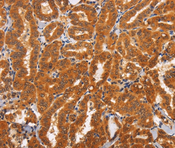
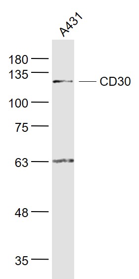
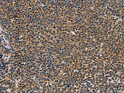
- Scientific DescriptionCD30 (Ki-1; TNF Receptor Superfamily Member 8) is a type I transmembrane glycoprotein of the TNF receptor superfamily. CD30 was originally identified as a cell surface antigen of Hodgkins and Reed-Sternberg cells using monoclonal antibody Ki-1. The ligand for CD30 is CD30L (CD153). The binding of CD30 to CD30L mediates pleiotropic effects including cell proliferation, activation, differentiation and apoptotic cell death. CD30 has a critical role in the pathophysiology of Hodgkins disease and other CD30+ lymphomas. CD30 acts as a costimulatory molecule in thymic negative selection. In addition to its expression on Hodgkins and Reed-Sternberg cells, CD30 is also found in some non-Hodgkins lymphomas (including Burkitts lymphomas), virus-infected T and B cells, and on normal T and B cells after activation. In T cells, CD30 expression is present on a subset of T cells that produce Th2-type cytokines and on CD4+/CD8+ thymocytes that co-express CD45RO and the IL4 receptor. Soluble form of CD30 (sCD30) serves as a marker reflecting Th2 immune response. TRAF2 and TRAF5 can interact with this receptor, and mediate the signal transduction that leads to the activation of NF-kappaB. CD30 is a positive regulator of apoptosis, and has been shown to limit the proliferative potential of autoreactive CD8 effector T cells and protect the body against autoimmunity. - Recombinant Antibody. Recognizes human CD30. Applications: ELISA, FACS, FUNC, ICC, WB. Clone: SH313-B5. Isotype: Human IgG1. Formulation: Liquid. In PBS. CD30 (Ki-1; TNF Receptor Superfamily Member 8) is a type I transmembrane glycoprotein of the TNF receptor superfamily. CD30 was originally identified as a cell surface antigen of Hodgkins and Reed-Sternberg cells using monoclonal antibody Ki-1. The ligand for CD30 is CD30L (CD153). The binding of CD30 to CD30L mediates pleiotropic effects including cell proliferation, activation, differentiation and apoptotic cell death. CD30 has a critical role in the pathophysiology of Hodgkins disease and other CD30+ lymphomas. CD30 acts as a costimulatory molecule in thymic negative selection. In addition to its expression on Hodgkins and Reed-Sternberg cells, CD30 is also found in some non-Hodgkins lymphomas (including Burkitts lymphomas), virus-infected T and B cells, and on normal T and B cells after activation. In T cells, CD30 expression is present on a subset of T cells that produce Th2-type cytokines and on CD4+/CD8+ thymocytes that co-express CD45RO and the IL4 receptor. Soluble form of CD30 (sCD30) serves as a marker reflecting Th2 immune response. TRAF2 and TRAF5 can interact with this receptor, and mediate the signal transduction that leads to the activation of NF-kappaB. CD30 is a positive regulator of apoptosis, and has been shown to limit the proliferative potential of autoreactive CD8 effector T cells and protect the body against autoimmunity.
- ReactivityHuman
- Storage Instruction-20°C,2°C to 8°C
- UNSPSC41116161
MSDS
Related products
Product group Antibodies
Anti-TNFRSF8 AntibodyA45177
ApplicationsImmunoHistoChemistry
ReactivityHuman
- SizePrice
Product group Antibodies

Anti-CD30 [Ki-3]Ab00474-1.1
ApplicationsFlow Cytometry, ImmunoFluorescence, ELISA, ImmunoHistoChemistry, Other Application
ReactivityHuman
TargetTNFRSF8
- SizePrice

Product group Antibodies
Anti-CD30/TNFRSF8 Antibody Picoband(r)A01225-2-CARRIER-FREE
ApplicationsWestern Blot, ImmunoHistoChemistry
ReactivityHuman, Mouse
TargetTNFRSF8
- SizePrice
Product group Antibodies
Anti-TNFRSF8 Antibody144-07651
ApplicationsWestern Blot, ImmunoHistoChemistry
ReactivityHuman, Mouse, Rat
TargetTNFRSF8
- SizePrice

Product group Antibodies
Anti-TNFRSF8 AntibodyAMAB91800
ApplicationsImmunoHistoChemistry
ReactivityHuman
TargetTNFRSF8
- SizePrice
Product group Antibodies
CD30 Polyclonal AntibodyBS-2495R
ApplicationsFlow Cytometry, ImmunoFluorescence, Western Blot, ELISA, ImmunoCytoChemistry, ImmunoHistoChemistry, ImmunoHistoChemistry Frozen, ImmunoHistoChemistry Paraffin
ReactivityHuman, Porcine, Rabbit
TargetTNFRSF8
- SizePrice
Product group Antibodies
TNFRSF8 AntibodyCSB-PA172042
ApplicationsELISA, ImmunoHistoChemistry
ReactivityHuman
TargetTNFRSF8
- SizePrice
Product group Antibodies
ApplicationsImmunoPrecipitation, Western Blot, ImmunoCytoChemistry, ImmunoHistoChemistry
TargetTNFRSF8
- SizePrice
Product group Antibodies

CD30 AntibodyLS-C403034
ApplicationsELISA, ImmunoHistoChemistry
ReactivityHuman
TargetTNFRSF8
- SizePrice