

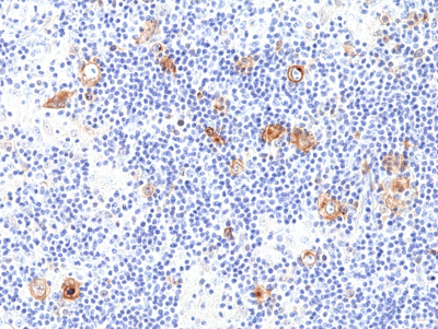
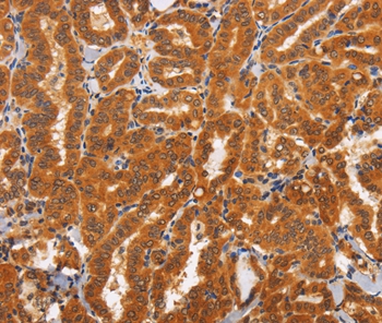
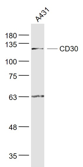
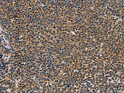
anti-CD30 Rabbit Monoclonal (RM425)
REV-31-1312-00
ApplicationsWestern Blot, ImmunoHistoChemistry
Product group Antibodies
ReactivityHuman
TargetTNFRSF8
Overview
- SupplierRevMAb Biosciences
- Product Nameanti-CD30 Rabbit Monoclonal (RM425)
- Delivery Days Customer2
- ApplicationsWestern Blot, ImmunoHistoChemistry
- CertificationResearch Use Only
- ClonalityMonoclonal
- Clone IDRM425
- Gene ID943
- Target nameTNFRSF8
- Target descriptionTNF receptor superfamily member 8
- Target synonymsCD30, D1S166E, Ki-1, tumor necrosis factor receptor superfamily member 8, CD30L receptor, Ki-1 antigen, cytokine receptor CD30, lymphocyte activation antigen CD30
- HostRabbit
- IsotypeIgG
- Protein IDP28908
- Protein NameTumor necrosis factor receptor superfamily member 8
- Scientific DescriptionCD30 (Ki-1, TNF Receptor Superfamily Member 8) is a type I transmembrane glycoprotein of the TNF receptor superfamily. CD30 was originally identified as a cell surface antigen of Hodgkins and Reed-Sternberg cells using monoclonal antibody Ki-1. The ligand for CD30 is CD30L (CD153). The binding of CD30 to CD30L mediates pleiotropic effects including cell proliferation, activation, differentiation, and apoptotic cell death. CD30 has a critical role in the pathophysiology of Hodgkins disease and other CD30+ lymphomas. CD30 acts as a costimulatory molecule in thymic negative selection. In addition to its expression on Hodgkins and Reed-Sternberg cells, CD30 is also found in some non-Hodgkins lymphomas (including Burkitts lymphomas), virus-infected T and B cells, and on normal T and B cells after activation. In T cells, CD30 expression is present on a subset of T cells that produce Th2-type cytokines and on CD4+/CD8+ thymocytes that co-express CD45RO and the IL4 receptor. Soluble form of CD30 (sCD30) serves as a marker reflecting Th2 immune response. TRAF2 and TRAF5 can interact with this receptor, and mediate the signal transduction that leads to the activation of NF-kappaB. CD30 is a positive regulator of apoptosis, and has been shown to limit the proliferative potential of autoreactive CD8 effector T cells and protect the body against autoimmunity. Two alternatively spliced transcript variants of this gene encoding distinct isoforms have been reported. CD30 is expressed by mononuclear cells in Hodgkins lymphoma, Reed Sternberg cells and most Anaplastic Large Cell Lymphomas (ALCL). CD30 is also expressed by embryonal carcinomas. - Recombinant Antibody. This antibody RM425 reacts to human CD30. Application: IHC, WB. Source: Rabbit. Liquid. 50% Glycerol/PBS with 1% BSA and 0.09% sodium azide. CD30 (Ki-1, TNF Receptor Superfamily Member 8) is a type I transmembrane glycoprotein of the TNF receptor superfamily. CD30 was originally identified as a cell surface antigen of Hodgkins and Reed-Sternberg cells using monoclonal antibody Ki-1. The ligand for CD30 is CD30L (CD153). The binding of CD30 to CD30L mediates pleiotropic effects including cell proliferation, activation, differentiation, and apoptotic cell death. CD30 has a critical role in the pathophysiology of Hodgkins disease and other CD30+ lymphomas. CD30 acts as a costimulatory molecule in thymic negative selection. In addition to its expression on Hodgkins and Reed-Sternberg cells, CD30 is also found in some non-Hodgkins lymphomas (including Burkitts lymphomas), virus-infected T and B cells, and on normal T and B cells after activation. In T cells, CD30 expression is present on a subset of T cells that produce Th2-type cytokines and on CD4+/CD8+ thymocytes that co-express CD45RO and the IL4 receptor. Soluble form of CD30 (sCD30) serves as a marker reflecting Th2 immune response. TRAF2 and TRAF5 can interact with this receptor, and mediate the signal transduction that leads to the activation of NF-kappaB. CD30 is a positive regulator of apoptosis, and has been shown to limit the proliferative potential of autoreactive CD8 effector T cells and protect the body against autoimmunity. Two alternatively spliced transcript variants of this gene encoding distinct isoforms have been reported. CD30 is expressed by mononuclear cells in Hodgkins lymphoma, Reed Sternberg cells and most Anaplastic Large Cell Lymphomas (ALCL). CD30 is also expressed by embryonal carcinomas.
- ReactivityHuman
- Storage Instruction-20°C,2°C to 8°C
- UNSPSC41116161
Datasheet
Related products
Product group Antibodies
Anti-TNFRSF8 AntibodyA45177
ApplicationsImmunoHistoChemistry
ReactivityHuman
- SizePrice
Product group Antibodies
anti-CD30 (human), mAb (rec.) (SH313-B5)AG-27B-6314
ApplicationsFunctional Assay, Flow Cytometry, Western Blot, ELISA, ImmunoCytoChemistry
ReactivityHuman
TargetTNFRSF8
- SizePrice
Product group Antibodies

Anti-CD30 [Ki-3]Ab00474-1.1
ApplicationsFlow Cytometry, ImmunoFluorescence, ELISA, ImmunoHistoChemistry, Other Application
ReactivityHuman
TargetTNFRSF8
- SizePrice

Product group Antibodies
Anti-CD30/TNFRSF8 Antibody Picoband(r)A01225-2-CARRIER-FREE
ApplicationsWestern Blot, ImmunoHistoChemistry
ReactivityHuman, Mouse
TargetTNFRSF8
- SizePrice
Product group Antibodies
Anti-TNFRSF8 Antibody144-07651
ApplicationsWestern Blot, ImmunoHistoChemistry
ReactivityHuman, Mouse, Rat
TargetTNFRSF8
- SizePrice

Product group Antibodies
Anti-TNFRSF8 AntibodyAMAB91800
ApplicationsImmunoHistoChemistry
ReactivityHuman
TargetTNFRSF8
- SizePrice
Product group Antibodies
CD30 Polyclonal AntibodyBS-2495R
ApplicationsFlow Cytometry, ImmunoFluorescence, Western Blot, ELISA, ImmunoCytoChemistry, ImmunoHistoChemistry, ImmunoHistoChemistry Frozen, ImmunoHistoChemistry Paraffin
ReactivityHuman, Porcine, Rabbit
TargetTNFRSF8
- SizePrice
Product group Antibodies
TNFRSF8 AntibodyCSB-PA172042
ApplicationsELISA, ImmunoHistoChemistry
ReactivityHuman
TargetTNFRSF8
- SizePrice
Product group Antibodies
ApplicationsImmunoPrecipitation, Western Blot, ImmunoCytoChemistry, ImmunoHistoChemistry
TargetTNFRSF8
- SizePrice