


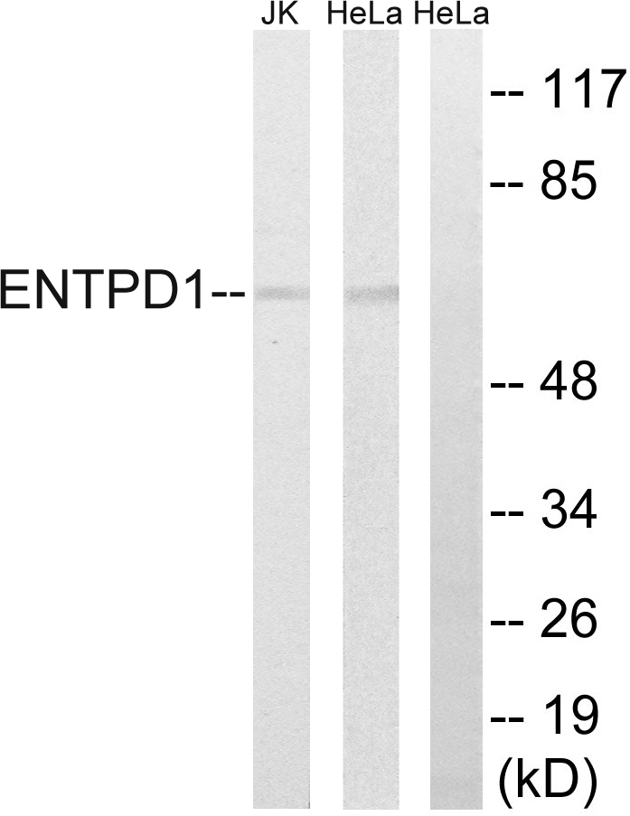
Figure 1. Western blot analysis of CD39/ENTPD1 using anti-CD39/ENTPD1 antibody (A03196-3). Electrophoresis was performed on a 5-20% SDS-PAGE gel at 70V (Stacking gel) / 90V (Resolving gel) for 2-3 hours. The sample well of each lane was loaded with 30 ug of sample under reducing conditions. Lane 1: human placenta tissue lysates. After electrophoresis, proteins were transferred to a nitrocellulose membrane at 150 mA for 50-90 minutes. Blocked the membrane with 5% non-fat milk/TBS for 1.5 hour at RT. The membrane was incubated with rabbit anti-CD39/ENTPD1 antigen affinity purified polyclonal antibody (Catalog # A03196-3) at 0.5 microg/mL overnight at 4°C, then washed with TBS-0.1%Tween 3 times with 5 minutes each and probed with a goat anti-rabbit IgG-HRP secondary antibody at a dilution of 1:5000 for 1.5 hour at RT. The signal is developed using an Enhanced Chemiluminescent detection (ECL) kit (Catalog # EK1002) with Tanon 5200 system. A specific band was detected for CD39/ENTPD1 at approximately 80 kDa. The expected band size for CD39/ENTPD1 is at 80 kDa.
. CD39/ENTPD1 was detected in a paraffin-embedded section of human gastric cancer tissue. Heat mediated antigen retrieval was performed in EDTA buffer (pH 8.0, epitope retrieval solution). The tissue section was blocked with 10% goat serum. The tissue section was then incubated with 2 microg/ml rabbit anti-CD39/ENTPD1 Antibody (A03196-3) overnight at 4°C. Biotinylated goat anti-rabbit IgG was used as secondary antibody and incubated for 30 minutes at 37°C. The tissue section was developed using Strepavidin-Biotin-Complex (SABC) (Catalog # SA1022) with DAB as the chromogen.")
. CD39/ENTPD1 was detected in a paraffin-embedded section of human liver cancer tissue. Heat mediated antigen retrieval was performed in EDTA buffer (pH 8.0, epitope retrieval solution). The tissue section was blocked with 10% goat serum. The tissue section was then incubated with 2 microg/ml rabbit anti-CD39/ENTPD1 Antibody (A03196-3) overnight at 4°C. Biotinylated goat anti-rabbit IgG was used as secondary antibody and incubated for 30 minutes at 37°C. The tissue section was developed using Strepavidin-Biotin-Complex (SABC) (Catalog # SA1022) with DAB as the chromogen.")
. CD39/ENTPD1 was detected in a paraffin-embedded section of human lung cancer tissue. Heat mediated antigen retrieval was performed in EDTA buffer (pH 8.0, epitope retrieval solution). The tissue section was blocked with 10% goat serum. The tissue section was then incubated with 2 microg/ml rabbit anti-CD39/ENTPD1 Antibody (A03196-3) overnight at 4°C. Biotinylated goat anti-rabbit IgG was used as secondary antibody and incubated for 30 minutes at 37°C. The tissue section was developed using Strepavidin-Biotin-Complex (SABC) (Catalog # SA1022) with DAB as the chromogen.")
. CD39/ENTPD1 was detected in a paraffin-embedded section of human ovarian cancer tissue. Heat mediated antigen retrieval was performed in EDTA buffer (pH 8.0, epitope retrieval solution). The tissue section was blocked with 10% goat serum. The tissue section was then incubated with 2 microg/ml rabbit anti-CD39/ENTPD1 Antibody (A03196-3) overnight at 4°C. Biotinylated goat anti-rabbit IgG was used as secondary antibody and incubated for 30 minutes at 37°C. The tissue section was developed using Strepavidin-Biotin-Complex (SABC) (Catalog # SA1022) with DAB as the chromogen.")
. CD39/ENTPD1 was detected in a paraffin-embedded section of human placenta tissue. Heat mediated antigen retrieval was performed in EDTA buffer (pH 8.0, epitope retrieval solution). The tissue section was blocked with 10% goat serum. The tissue section was then incubated with 2 microg/ml rabbit anti-CD39/ENTPD1 Antibody (A03196-3) overnight at 4°C. Biotinylated goat anti-rabbit IgG was used as secondary antibody and incubated for 30 minutes at 37°C. The tissue section was developed using Strepavidin-Biotin-Complex (SABC) (Catalog # SA1022) with DAB as the chromogen.")
. CD39/ENTPD1 was detected in a paraffin-embedded section of human colon cancer tissue. Heat mediated antigen retrieval was performed in EDTA buffer (pH 8.0, epitope retrieval solution). The tissue section was blocked with 10% goat serum. The tissue section was then incubated with 4 microg/mL rabbit anti-CD39/ENTPD1 Antibody (A03196-3) overnight at 4°C. Biotin conjugated goat anti-rabbit IgG (BA1003) was used as secondary antibody and incubated for 30 minutes at 37°C. The tissue section was developed using DyLight®488 Conjugated Avidin (BA1128). The section was counterstained with DAPI. Visualize using a fluorescence microscope and filter sets appropriate for the label used.")
. Overlay histogram showing THP-1 cells stained with A03196-3 (Blue line). The cells were fixed with 4% paraformaldehyde and blocked with 10% normal goat serum. And then incubated with rabbit anti-CD39/ENTPD1 Antibody (A03196-3, 1 microg/1x106 cells) for 30 min at 20°C. DyLight®488 conjugated goat anti-rabbit IgG (BA1127, 5-10 microg/1x106 cells) was used as secondary antibody for 30 minutes at 20°C. Isotype control antibody (Green line) was rabbit IgG (1 microg/1x106) used under the same conditions. Unlabelled sample without incubation with primary antibody and secondary antibody (Red line) was used as a blank control.")
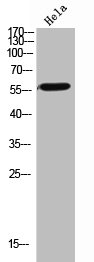
Figure 1. Western blot analysis of CD39/ENTPD1 using anti-CD39/ENTPD1 antibody (A03196-3). Electrophoresis was performed on a 5-20% SDS-PAGE gel at 70V (Stacking gel) / 90V (Resolving gel) for 2-3 hours. The sample well of each lane was loaded with 30 ug of sample under reducing conditions. Lane 1: human placenta tissue lysates. After electrophoresis, proteins were transferred to a nitrocellulose membrane at 150 mA for 50-90 minutes. Blocked the membrane with 5% non-fat milk/TBS for 1.5 hour at RT. The membrane was incubated with rabbit anti-CD39/ENTPD1 antigen affinity purified polyclonal antibody (Catalog # A03196-3) at 0.5 microg/mL overnight at 4°C, then washed with TBS-0.1%Tween 3 times with 5 minutes each and probed with a goat anti-rabbit IgG-HRP secondary antibody at a dilution of 1:5000 for 1.5 hour at RT. The signal is developed using an Enhanced Chemiluminescent detection (ECL) kit (Catalog # EK1002) with Tanon 5200 system. A specific band was detected for CD39/ENTPD1 at approximately 80 kDa. The expected band size for CD39/ENTPD1 is at 80 kDa.
Anti-CD39/ENTPD1 Antibody Picoband(r)
A03196-3-CARRIER-FREE
ApplicationsFlow Cytometry, ImmunoFluorescence, Western Blot, ELISA, ImmunoHistoChemistry
Product group Antibodies
ReactivityHuman
TargetENTPD1
Overview
- SupplierBoster Bio
- Product NameAnti-CD39/ENTPD1 Antibody Picoband(r)
- Delivery Days Customer9
- ApplicationsFlow Cytometry, ImmunoFluorescence, Western Blot, ELISA, ImmunoHistoChemistry
- CertificationResearch Use Only
- ClonalityPolyclonal
- Concentration500 ug/ml
- Gene ID953
- Target nameENTPD1
- Target descriptionectonucleoside triphosphate diphosphohydrolase 1
- Target synonymsATP-DPH, ATPDase, CD39, NTPDase-1, SPG64, ectonucleoside triphosphate diphosphohydrolase 1, ATP diphosphohydrolase, CD39 antigen, NTPDase1, ecto-ATP diphosphohydrolase 1, ecto-ATPDase 1, ecto-ATPase 1, ecto-apyrase, lymphoid cell activation antigen, nucleoside triphosphate diphosphohydrolase 1
- HostRabbit
- IsotypeIgG
- Protein IDP49961
- Protein NameEctonucleoside triphosphate diphosphohydrolase 1
- Scientific DescriptionBoster Bio Anti-CD39/ENTPD1 Antibody Picoband® catalog # A03196-3. Tested in ELISA, Flow Cytometry, IF, IHC, WB applications. This antibody reacts with Human. The brand Picoband indicates this is a premium antibody that guarantees superior quality, high affinity, and strong signals with minimal background in Western blot applications. Only our best-performing antibodies are designated as Picoband, ensuring unmatched performance.
- ReactivityHuman
- Storage Instruction-20°C,2°C to 8°C
- UNSPSC12352203
Related products
Product group Antibodies

Anti-CD39 [R22]Ab02890-10.0
ApplicationsFlow Cytometry
ReactivityHuman
TargetENTPD1
- SizePrice
Product group Antibodies
Anti-ENTPD1 AntibodyA100576
ApplicationsWestern Blot, ELISA
ReactivityHuman
- SizePrice
Product group Antibodies
Anti-ENTPD1 Antibody144-03778
ApplicationsWestern Blot
ReactivityHuman, Mouse, Rat
TargetENTPD1
- SizePrice

Product group Antibodies

CD39 Antibody (clone A1, FITC)LS-C811814
ApplicationsFlow Cytometry
ReactivityHuman
TargetENTPD1
- SizePrice

Product group Antibodies
CD39/ENTPD1 Recombinant AntibodyBSM-54043R
ApplicationsImmunoFluorescence, ImmunoHistoChemistry, ImmunoHistoChemistry Frozen, ImmunoHistoChemistry Paraffin
ReactivityHuman
TargetENTPD1
- SizePrice
Product group Antibodies
ENTPD1 AntibodyCSB-PA006195
ApplicationsWestern Blot, ELISA
ReactivityHuman, Mouse, Rat
TargetENTPD1
- SizePrice
Product group Antibodies
Entpd1 Polyclonal AntibodyCAC11591
ApplicationsImmunoFluorescence, Western Blot, ELISA, ImmunoHistoChemistry
TargetENTPD1
- SizePrice
Product group Antibodies

ApplicationsFlow Cytometry
ReactivityHuman
- SizePrice