

CD39 Antibody (APC)
ABX140487
ApplicationsFlow Cytometry
Product group Antibodies
Overview
- SupplierAbbexa
- Product NameCD39 Antibody (APC)
- Delivery Days Customer12
- ApplicationsFlow Cytometry
- CertificationResearch Use Only
- ClonalityMonoclonal
- ConjugateAPC (Allophycocyanin)
- HostMouse
- UNSPSC12352203
Related products
Product group Antibodies

Anti-CD39 [R22]Ab02890-10.0
ApplicationsFlow Cytometry
ReactivityHuman
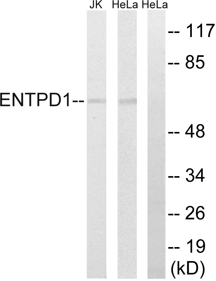
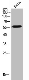
TargetENTPD1
- SizePrice
Product group Antibodies
Anti-ENTPD1 AntibodyA100576
ApplicationsWestern Blot, ELISA
ReactivityHuman
- SizePrice

Product group Antibodies
Anti-CD39/ENTPD1 Antibody Picoband(r)A03196-3-CARRIER-FREE
ApplicationsFlow Cytometry, ImmunoFluorescence, Western Blot, ELISA, ImmunoHistoChemistry
ReactivityHuman
TargetENTPD1
- SizePrice
Product group Antibodies
Anti-ENTPD1 Antibody144-03778
ApplicationsWestern Blot
ReactivityHuman, Mouse, Rat
TargetENTPD1
- SizePrice
Product group Antibodies

CD39 Antibody (clone A1, FITC)LS-C811814
ApplicationsFlow Cytometry
ReactivityHuman
TargetENTPD1
- SizePrice

Product group Antibodies
CD39/ENTPD1 Recombinant AntibodyBSM-54043R
ApplicationsImmunoFluorescence, ImmunoHistoChemistry, ImmunoHistoChemistry Frozen, ImmunoHistoChemistry Paraffin
ReactivityHuman
TargetENTPD1
- SizePrice
Product group Antibodies
ENTPD1 AntibodyCSB-PA006195
ApplicationsWestern Blot, ELISA
ReactivityHuman, Mouse, Rat
TargetENTPD1
- SizePrice
Product group Antibodies
Entpd1 Polyclonal AntibodyCAC11591
ApplicationsImmunoFluorescence, Western Blot, ELISA, ImmunoHistoChemistry
TargetENTPD1
- SizePrice
Product group Antibodies

ApplicationsFlow Cytometry
ReactivityHuman
- SizePrice