


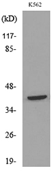
Figure 1. Western blot analysis of CD48 using anti-CD48 antibody (A03281-1). Electrophoresis was performed on a 5-20% SDS-PAGE gel at 70V (Stacking gel) / 90V (Resolving gel) for 2-3 hours. The sample well of each lane was loaded with 50ug of sample under reducing conditions. lane 1: CEM whole cell lysates. After Electrophoresis, proteins were transferred to a Nitrocellulose membrane at 150mA for 50-90 minutes. Blocked the membrane with 5% Non-fat Milk/ TBS for 1.5 hour at RT. The membrane was incubated with rabbit anti-CD48 antigen affinity purified polyclonal antibody (Catalog # A03281-1) at 0.5 microg/mL overnight at 4°C, then washed with TBS-0.1%Tween 3 times with 5 minutes each and probed with a goat anti-rabbit IgG-HRP secondary antibody at a dilution of 1:10000 for 1.5 hour at RT. The signal is developed using an Enhanced Chemiluminescent detection (ECL) kit (Catalog # EK1002) with Tanon 5200 system. A specific band was detected for CD48 at approximately 39KD. The expected band size for CD48 is at 28KD.
Figure 1. Western blot analysis of CD48 using anti-CD48 antibody (A03281-1). Electrophoresis was performed on a 5-20% SDS-PAGE gel at 70V (Stacking gel) / 90V (Resolving gel) for 2-3 hours. The sample well of each lane was loaded with 50ug of sample under reducing conditions. lane 1: CEM whole cell lysates. After Electrophoresis, proteins were transferred to a Nitrocellulose membrane at 150mA for 50-90 minutes. Blocked the membrane with 5% Non-fat Milk/ TBS for 1.5 hour at RT. The membrane was incubated with rabbit anti-CD48 antigen affinity purified polyclonal antibody (Catalog # A03281-1) at 0.5 microg/mL overnight at 4°C, then washed with TBS-0.1%Tween 3 times with 5 minutes each and probed with a goat anti-rabbit IgG-HRP secondary antibody at a dilution of 1:10000 for 1.5 hour at RT. The signal is developed using an Enhanced Chemiluminescent detection (ECL) kit (Catalog # EK1002) with Tanon 5200 system. A specific band was detected for CD48 at approximately 39KD. The expected band size for CD48 is at 28KD.
Anti-CD48 Antibody Picoband(r)
A03281-1-CARRIER-FREE
ApplicationsWestern Blot
Product group Antibodies
ReactivityHuman
TargetCD48
Overview
- SupplierBoster Bio
- Product NameAnti-CD48 Antibody Picoband(r)
- Delivery Days Customer9
- ApplicationsWestern Blot
- CertificationResearch Use Only
- ClonalityPolyclonal
- Concentration500 ug/ml
- Gene ID962
- Target nameCD48
- Target descriptionCD48 molecule
- Target synonymsBCM1, BLAST, BLAST1, MEM-102, SLAMF2, hCD48, mCD48, CD48 antigen, B-lymphocyte activation marker BLAST-1, BCM1 surface antigen, CD48 antigen (B-cell membrane protein), SLAM family member 2, TCT.1, leukocyte antigen MEM-102, signaling lymphocytic activation molecule 2
- HostRabbit
- IsotypeIgG
- Protein IDP09326
- Protein NameCD48 antigen
- Scientific DescriptionBoster Bio Anti-CD48 Antibody Picoband® catalog # A03281-1. Tested in WB applications. This antibody reacts with Human. The brand Picoband indicates this is a premium antibody that guarantees superior quality, high affinity, and strong signals with minimal background in Western blot applications. Only our best-performing antibodies are designated as Picoband, ensuring unmatched performance.
- ReactivityHuman
- Storage Instruction-20°C,2°C to 8°C
- UNSPSC12352203
Related products
Product group Antibodies
Anti-CD48 AntibodyA101511
ApplicationsWestern Blot, ELISA
ReactivityHuman
- SizePrice
Product group Antibodies
Anti-CD48 Antibody188-10813
ApplicationsImmunoFluorescence
ReactivityHuman
TargetCD48
- SizePrice

Product group Antibodies

CD48 Antibody (clone HM48-1)LS-C770051
ApplicationsFlow Cytometry, ImmunoPrecipitation, ImmunoHistoChemistry
ReactivityMouse
TargetCD48
- SizePrice
Product group Antibodies
CD48 Recombinant Antibody, Biotin ConjugatedBSM-61753R-BIOTIN
ApplicationsImmunoPrecipitation, Western Blot, ImmunoHistoChemistry, ImmunoHistoChemistry Frozen, ImmunoHistoChemistry Paraffin
ReactivityHuman
TargetCD48
- SizePrice
Product group Antibodies
ApplicationsImmunoPrecipitation, Western Blot, ImmunoCytoChemistry, ImmunoHistoChemistry
TargetCD48
- SizePrice
Product group Antibodies
CD48 AntibodyCSB-PA004941ESR1HU
ApplicationsELISA, ImmunoHistoChemistry
ReactivityHuman
TargetCD48
- SizePrice
![FACS analysis of human peripheral blood using GTX78302 CD48 antibody [MEM-102] (FITC). Antibody amount : 20 μl reagent / 100 μl of peripheral whole blood](https://www.genetex.com/upload/website/prouct_img/normal/GTX78302/GTX78302_20191025_AP_006_283_w_23061322_457.webp)
Product group Antibodies
CD48 antibody [MEM-102] (FITC)GTX78302
ApplicationsFlow Cytometry
ReactivityHuman, Primate
TargetCD48
- SizePrice

Product group Antibodies
Anti-CD48 AntibodyHPA055146
ApplicationsImmunoCytoChemistry
ReactivityHuman
TargetCD48
- SizePrice