

Anti-CD99 [12E7]
Ab02834-1.1
ApplicationsFlow Cytometry, ImmunoFluorescence, Western Blot, ImmunoCytoChemistry, ImmunoHistoChemistry, RadioImmunoAssay
Product group Antibodies
ReactivityHuman
TargetCD99
Overview
- SupplierAbsolute Antibody
- Product NameAnti-CD99 [12E7]
- Delivery Days Customer7
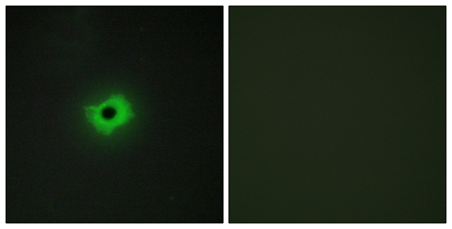
- Application Supplier NoteThe original mouse IgG1 version of this antibody was used for the indirect immunofluorescence staining of a fixed bone marrow smear from a patient with T-cell leukemia. The binding specificity of this antibody towards CD99 was tested using radioimmunoassay (PMID: 316541). This antibody was able to identify 28 kDa component present on lymphocytes, red blood cells and platelets in a western blot assay (PMID: 2447875). This antibody was used in the immunocytochemical studies of Ewings sarcoma and peripheral neuroectodermal tumour (PMID: 8314240). This antibody was also used in the immunohistochemical studies of formalin-fixed, paraffin-embedded lymphoblastic lymphoma (LyL)/acute lymphoblastic leukemia (ALL) samples collected from 24 patients (PMID: 9343323). This antibody was also used in a study relating to the cell surface expression MIC2 antigen on human leukocytes in bone marrow (BM), thymus, and peripheral blood (PB) using multiparameter flow cytometry and cell sorting (PMID: 7506950).
- ApplicationsFlow Cytometry, ImmunoFluorescence, Western Blot, ImmunoCytoChemistry, ImmunoHistoChemistry, RadioImmunoAssay
- Applications SupplierIF; RIA; WB; ICC; IHC; FC
- CertificationResearch Use Only
- ClonalityMonoclonal
- Clone ID12E7
- Gene ID4267
- Target nameCD99
- Target descriptionCD99 molecule (Xg blood group)
- Target synonymsHBA71, MIC2, MIC2X, MIC2Y, MSK5X, CD99 antigen, E2 antigen, MIC2 (monoclonal antibody 12E7), T-cell surface glycoprotein E2, antigen identified by monoclonal 12E7, Y homolog, antigen identified by monoclonal antibodies 12E7, F21 and O13, cell surface antigen 12E7, cell surface antigen HBA-71, cell surface antigen O13, surface antigen MIC2
- HostMouse
- IsotypeIgG1
- Protein IDP14209
- Protein NameCD99 antigen
- ReactivityHuman
- Reactivity SupplierHuman
- Reactivity Supplier NoteThe original antibody was generated by immunizing BALB/c mice intraperitoneally with glutaraldehyde-fixed human acute lymphocytic leukemia (ALL) cells obtained from peripheral blood of a child.
- Storage Instruction-20°C,2°C to 8°C
- UNSPSC41116161
Related products
Product group Antibodies
Anti-CD99 AntibodyA101498
ApplicationsImmunoFluorescence, ELISA
ReactivityHuman
- SizePrice
Product group Antibodies
Anti-CD99 Antibody144-02028
ApplicationsWestern Blot
ReactivityHuman
TargetCD99
- SizePrice

Product group Antibodies
Anti-CD99 AntibodyAMAB91823
ApplicationsImmunoHistoChemistry
ReactivityHuman
TargetCD99
- SizePrice
Product group Antibodies

CD99 AntibodyLS-C831952
ApplicationsELISA, ImmunoHistoChemistry
ReactivityHuman
TargetCD99
- SizePrice

Product group Antibodies
Anti-CD99 Antibody Picoband(r)A01724-2-CARRIER-FREE
ApplicationsWestern Blot
ReactivityHuman
TargetCD99
- SizePrice
Product group Antibodies
ApplicationsFlow Cytometry, Western Blot
ReactivityHuman
TargetCD99
- SizePrice
Product group Antibodies
CD99 AntibodyCSB-PA010191
ApplicationsImmunoFluorescence, ELISA
ReactivityHuman
TargetCD99
- SizePrice
Product group Antibodies
ApplicationsImmunoPrecipitation, Western Blot, ImmunoCytoChemistry, ImmunoHistoChemistry
TargetCD99
- SizePrice
Product group Antibodies
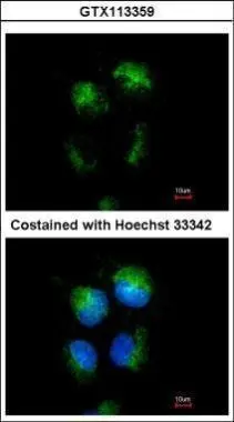
CD99 antibodyGTX113359
ApplicationsImmunoFluorescence, Western Blot, ImmunoCytoChemistry, ImmunoHistoChemistry, ImmunoHistoChemistry Paraffin
ReactivityHuman
TargetCD99
- SizePrice
Product group Antibodies
Anti-SARS-CoV-2 NSP2 AntibodyCAB20280
ApplicationsWestern Blot, ELISA
TargetCD99
- SizePrice