

Anti-CDC73 (Center) Antibody
102-27676
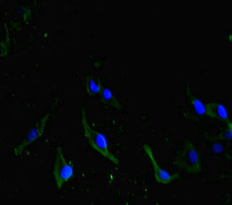
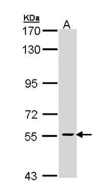
ApplicationsFlow Cytometry, ImmunoFluorescence, Western Blot, ImmunoHistoChemistry, ImmunoHistoChemistry Paraffin
Product group Antibodies
TargetCDC73
Overview
- SupplierRayBiotech
- Product NameAnti-CDC73 (Center) Antibody
- Delivery Days Customer16
- ApplicationsFlow Cytometry, ImmunoFluorescence, Western Blot, ImmunoHistoChemistry, ImmunoHistoChemistry Paraffin
- CertificationResearch Use Only
- ClonalityPolyclonal
- Clone IDRB23239
- Gene ID79577
- Target nameCDC73
- Target descriptioncell division cycle 73
- Target synonymsC1orf28, FIHP, HPTJT, HRPT1, HRPT2, HYX, parafibromin, Familial isolated hyperparathyroidism, Paf1/RNA polymerase II complex component, cell division cycle 73 Paf1/RNA polymerase II complex component-like protein, cell division cycle 73, Paf1/RNA polymerase II complex component, homolog, cell division cycle protein 73 homolog, hyperparathyroidism 2 protein
- HostRabbit
- Protein IDQ4V8C8
- Protein NameParafibromin
- Scientific DescriptionRabbit Anti-CDC73 (Center) Antibody, 400 microl
- Storage Instruction-20°C
- UNSPSC12352203
Related products
Product group Antibodies

Anti-CDC73 [RAB-C146]Ab01755-1.1
ApplicationsFlow Cytometry, ImmunoFluorescence, ImmunoPrecipitation
ReactivityHuman
TargetCDC73
- SizePrice

Product group Antibodies
Anti-CDC73 AntibodyHPA030772
ApplicationsImmunoCytoChemistry
ReactivityHuman
TargetCDC73
- SizePrice
Product group Antibodies
CDC73 AntibodyCSB-PA750783LA01HU
ApplicationsImmunoFluorescence, ELISA
ReactivityHuman
TargetCDC73
- SizePrice
Product group Antibodies

CDC73 / Parafibromin AntibodyLS-C331037
ApplicationsFlow Cytometry, ImmunoFluorescence, Western Blot, ImmunoHistoChemistry
ReactivityHuman, Mouse
TargetCDC73
- SizePrice

Product group Antibodies
Anti-HRPT2/CDC73 Antibody Picoband(r)PB9532-CARRIER-FREE
ApplicationsWestern Blot
ReactivityHuman
TargetCDC73
- SizePrice
Product group Antibodies
CDC73 antibody [N1C1]GTX110280
ApplicationsWestern Blot
ReactivityHuman
TargetCDC73
- SizePrice
Product group Antibodies
ApplicationsFlow Cytometry, Western Blot, ImmunoCytoChemistry
ReactivityHuman
TargetCDC73
- SizePrice