

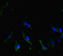
Immunofluorescent analysis of Hela cells using CSB-PA750783LA01HU at dilution of 1: 100 and Alexa Fluor 488-congugated AffiniPure Goat Anti-Rabbit IgG(H+L)
Immunofluorescent analysis of Hela cells using CSB-PA750783LA01HU at dilution of 1: 100 and Alexa Fluor 488-congugated AffiniPure Goat Anti-Rabbit IgG(H+L)
CDC73 Antibody
CSB-PA750783LA01HU
ApplicationsImmunoFluorescence, ELISA
Product group Antibodies
ReactivityHuman
TargetCDC73
Overview
- SupplierCusabio
- Product NameCDC73 Antibody
- Delivery Days Customer20
- ApplicationsImmunoFluorescence, ELISA
- CertificationResearch Use Only
- ClonalityPolyclonal
- ConjugateUnconjugated
- Gene ID79577
- Target nameCDC73
- Target descriptioncell division cycle 73
- Target synonymsC1orf28, FIHP, HPTJT, HRPT1, HRPT2, HYX, parafibromin, Familial isolated hyperparathyroidism, Paf1/RNA polymerase II complex component, cell division cycle 73 Paf1/RNA polymerase II complex component-like protein, cell division cycle 73, Paf1/RNA polymerase II complex component, homolog, cell division cycle protein 73 homolog, hyperparathyroidism 2 protein
- HostRabbit
- IsotypeIgG
- Protein IDQ6P1J9
- Protein NameParafibromin
- Scientific DescriptionTumor suppressor probably involved in transcriptional and post-transcriptional control pathways. May be involved in cell cycle progression through the regulation of cyclin D1/PRAD1 expression. Component of the PAF1 complex (PAF1C) which has multiple functions during transcription by RNA polymerase II and is implicated in regulation of development and maintenance of embryonic stem cell pluripotency. PAF1C associates with RNA polymerase II through interaction with POLR2A CTD non-phosphorylated and Ser-2- and Ser-5-phosphorylated forms and is involved in transcriptional elongation, acting both indepentently and synergistically with TCEA1 and in cooperation with the DSIF complex and HTATSF1. PAF1C is required for transcription of Hox and Wnt target genes. PAF1C is involved in hematopoiesis and stimulates transcriptional activity of KMT2A/MLL1; it promotes leukemogenesis through association with KMT2A/MLL1-rearranged oncoproteins, such as KMT2A/MLL1-MLLT3/AF9 and KMT2A/MLL1-MLLT1/ENL. PAF1C is involved in histone modifications such as ubiquitination of histone H2B and methylation on histone H3 Lys-4 (H3K4me3). PAF1C recruits the RNF20/40 E3 ubiquitin-protein ligase complex and the E2 enzyme UBE2A or UBE2B to chromatin which mediate monoubiquitination of Lys-120 of histone H2B (H2BK120ub1); UB2A/B-mediated H2B ubiquitination is proposed to be coupled to transcription. PAF1C is involved in mRNA 3 end formation probably through association with cleavage and poly(A) factors. In case of infection by influenza A strain H3N2, PAF1C associates with viral NS1 protein, thereby regulating gene transcription. Connects PAF1C with the cleavage and polyadenylation specificity factor (CPSF) complex and the cleavage stimulation factor (CSTF) complex, and with Wnt signaling. Involved in polyadenylation of mRNA precursors.
- ReactivityHuman
- Storage Instruction-20°C or -80°C
- UNSPSC41116161
Related products
Product group Antibodies

Anti-CDC73 [RAB-C146]Ab01755-1.1
ApplicationsFlow Cytometry, ImmunoFluorescence, ImmunoPrecipitation
ReactivityHuman
TargetCDC73
- SizePrice

Product group Antibodies
Anti-CDC73 AntibodyHPA030772
ApplicationsImmunoCytoChemistry
ReactivityHuman
TargetCDC73
- SizePrice
Product group Antibodies

CDC73 / Parafibromin AntibodyLS-C331037
ApplicationsFlow Cytometry, ImmunoFluorescence, Western Blot, ImmunoHistoChemistry
ReactivityHuman, Mouse
TargetCDC73
- SizePrice

Product group Antibodies
Anti-HRPT2/CDC73 Antibody Picoband(r)PB9532-CARRIER-FREE
ApplicationsWestern Blot
ReactivityHuman
TargetCDC73
- SizePrice
Product group Antibodies
CDC73 antibody [N1C1]GTX110280
ApplicationsWestern Blot
ReactivityHuman
TargetCDC73
- SizePrice
Product group Antibodies
ApplicationsFlow Cytometry, Western Blot, ImmunoCytoChemistry
ReactivityHuman
TargetCDC73
- SizePrice
Product group Antibodies
Anti-CDC73 (Center) Antibody102-27676
ApplicationsFlow Cytometry, ImmunoFluorescence, Western Blot, ImmunoHistoChemistry, ImmunoHistoChemistry Paraffin
TargetCDC73
- SizePrice