

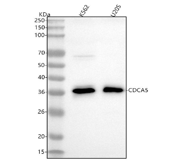
Figure 1. Western blot analysis of CDCA5 using anti-CDCA5 antibody (M05043). Electrophoresis was performed on a 5-20% SDS-PAGE gel at 70V (Stacking gel) / 90V (Resolving gel) for 2-3 hours. The sample well of each lane was loaded with 30 ug of sample under reducing conditions. Lane 1: human K562 whole cell lysates, Lane 2: human U2OS whole cell lysates. After electrophoresis, proteins were transferred to a nitrocellulose membrane at 150 mA for 50-90 minutes. Blocked the membrane with 5% non-fat milk/TBS for 1.5 hour at RT. The membrane was incubated with rabbit anti-CDCA5 antigen affinity purified monoclonal antibody (Catalog # M05043) at 1:500 overnight at 4°C, then washed with TBS-0.1%Tween 3 times with 5 minutes each and probed with a goat anti-rabbit IgG-HRP secondary antibody at a dilution of 1:500 for 1.5 hour at RT. The signal is developed using an Enhanced Chemiluminescent detection (ECL) kit (Catalog # EK1002) with Tanon 5200 system. A specific band was detected for CDCA5 at approximately 35 kDa. The expected band size for CDCA5 is at 28 kDa.


Figure 1. Western blot analysis of CDCA5 using anti-CDCA5 antibody (M05043). Electrophoresis was performed on a 5-20% SDS-PAGE gel at 70V (Stacking gel) / 90V (Resolving gel) for 2-3 hours. The sample well of each lane was loaded with 30 ug of sample under reducing conditions. Lane 1: human K562 whole cell lysates, Lane 2: human U2OS whole cell lysates. After electrophoresis, proteins were transferred to a nitrocellulose membrane at 150 mA for 50-90 minutes. Blocked the membrane with 5% non-fat milk/TBS for 1.5 hour at RT. The membrane was incubated with rabbit anti-CDCA5 antigen affinity purified monoclonal antibody (Catalog # M05043) at 1:500 overnight at 4°C, then washed with TBS-0.1%Tween 3 times with 5 minutes each and probed with a goat anti-rabbit IgG-HRP secondary antibody at a dilution of 1:500 for 1.5 hour at RT. The signal is developed using an Enhanced Chemiluminescent detection (ECL) kit (Catalog # EK1002) with Tanon 5200 system. A specific band was detected for CDCA5 at approximately 35 kDa. The expected band size for CDCA5 is at 28 kDa.
Anti-CDCA5/Sororin Rabbit Monoclonal Antibody
M05043
ApplicationsFlow Cytometry, ImmunoFluorescence, Western Blot, ImmunoCytoChemistry, ImmunoHistoChemistry
Product group Antibodies
ReactivityHuman
TargetCDCA5
Overview
- SupplierBoster Bio
- Product NameAnti-CDCA5/Sororin Rabbit Monoclonal Antibody
- Delivery Days Customer9
- ApplicationsFlow Cytometry, ImmunoFluorescence, Western Blot, ImmunoCytoChemistry, ImmunoHistoChemistry
- CertificationResearch Use Only
- ClonalityMonoclonal
- Clone IDIAH-3
- Gene ID113130
- Target nameCDCA5
- Target descriptioncell division cycle associated 5
- Target synonymsSORORIN, sororin, cell division cycle-associated protein 5, p35
- HostRabbit
- IsotypeIgG
- Protein IDQ96FF9
- Protein NameSororin
- Scientific DescriptionBoster Bio Anti-CDCA5/Sororin Rabbit Monoclonal Antibody catalog # M05043. Tested in WB, IHC, ICC/IF, Flow Cytometry applications. This antibody reacts with Human.
- ReactivityHuman
- Storage Instruction-20°C
- UNSPSC12352203
Datasheet
MSDS
Related products
Product group Antibodies
Anti-CDCA5 AntibodyA89350
ApplicationsWestern Blot
ReactivityHuman, Mouse, Rat
- SizePrice

Product group Antibodies
Anti-CDCA5 AntibodyHPA023691
ApplicationsWestern Blot, ImmunoCytoChemistry, ImmunoHistoChemistry
ReactivityHuman
TargetCDCA5
- SizePrice
Product group Antibodies

CDCA5 / Sororin AntibodyLS-C664515
ApplicationsWestern Blot, ELISA
ReactivityHuman
TargetCDCA5
- SizePrice
Product group Antibodies
CDCA5 Polyclonal AntibodyBS-7717R
ApplicationsFlow Cytometry, ImmunoFluorescence, Western Blot, ELISA, ImmunoCytoChemistry, ImmunoHistoChemistry, ImmunoHistoChemistry Frozen, ImmunoHistoChemistry Paraffin
ReactivityBovine, Canine, Equine, Human, Mouse, Porcine, Rabbit, Rat
TargetCDCA5
- SizePrice
Product group Antibodies
CDCA5 antibody, InternalGTX44862
ApplicationsWestern Blot
ReactivityHuman
TargetCDCA5
- SizePrice
Product group Antibodies
Anti-CDCA5 Antibody144-64522
ApplicationsWestern Blot
ReactivityHuman, Mouse, Rat
TargetCDCA5
- SizePrice