


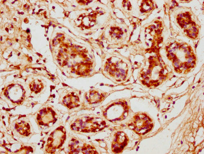
Immunohistochemical staining of human kidney shows strong nuclear and cytoplasmic positivity in cells in tubules.

Immunohistochemical staining of human kidney shows strong nuclear and cytoplasmic positivity in cells in tubules.
Anti-CDK3 Antibody
HPA007420
ApplicationsImmunoCytoChemistry, ImmunoHistoChemistry
Product group Antibodies
ReactivityHuman
TargetCDK3
Overview
- SupplierAtlas Antibodies
- Product NameAnti-CDK3 Antibody
- Delivery Days Customer4
- ApplicationsImmunoCytoChemistry, ImmunoHistoChemistry
- CertificationResearch Use Only
- ClonalityPolyclonal
- ConjugateUnconjugated
- Gene ID1018
- Target nameCDK3
- Target descriptioncyclin dependent kinase 3
- Target synonymscyclin-dependent kinase 3, cell division protein kinase 3
- HostRabbit
- IsotypeIgG
- Protein IDQ00526
- Protein NameCyclin-dependent kinase 3
- Scientific DescriptionRecombinant Protein Epitope Signature Tag (PrEST) antigen sequence
- ReactivityHuman
- Storage Instruction-20°C,2°C to 8°C
- UNSPSC41116161
Datasheet
MSDS
Related products
Product group Antibodies
CDK3 AntibodyCSB-PA005064LA01HU
ApplicationsELISA, ImmunoHistoChemistry
ReactivityHuman
TargetCDK3
- SizePrice

Product group Antibodies
ApplicationsWestern Blot
ReactivityHuman, Mouse, Rat
TargetCDK3
- SizePrice
Product group Antibodies
Anti-CDK3 AntibodyA28264
ApplicationsWestern Blot
ReactivityHuman, Mouse, Rat
- SizePrice
Product group Antibodies

CDK3 Antibody (N-Terminus)LS-C353710
ApplicationsWestern Blot
ReactivityHuman, Mouse, Rat
TargetCDK3
- SizePrice
Product group Antibodies
CDK3 Polyclonal AntibodyBS-1680R
ApplicationsImmunoFluorescence, Western Blot, ELISA, ImmunoCytoChemistry, ImmunoHistoChemistry, ImmunoHistoChemistry Frozen, ImmunoHistoChemistry Paraffin
ReactivityBovine, Canine, Chicken, Equine, Guinea Pig, Human, Mouse, Porcine, Rat
TargetCDK3
- SizePrice
Product group Antibodies
Anti-Cdk3 Antibody107-10507
ApplicationsWestern Blot
ReactivityHuman
TargetCDK3
- SizePrice
Product group Antibodies
CDK3 antibody [N1C2]GTX105047
ApplicationsWestern Blot
ReactivityHuman
TargetCDK3
- SizePrice
Product group Antibodies
CDK3 Polyclonal AntibodyCAC13006
ApplicationsELISA, ImmunoHistoChemistry
TargetCDK3
- SizePrice