


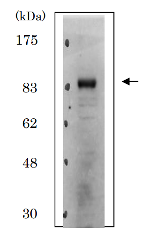
Figure 1. Western blot analysis of DTL using anti-DTL antibody (M01255). Electrophoresis was performed on a 5-20% SDS-PAGE gel at 70V (Stacking gel) / 90V (Resolving gel) for 2-3 hours. The sample well of each lane was loaded with 30 ug of sample under reducing conditions. Lane 1: human 293T whole cell lysates, Lane 2: human SH-SY5Y whole cell lysates, Lane 3: human K562 whole cell lysates, Lane 4: rat testis tissue lysates, Lane 5: rat thymus tissue lysates, Lane 6: mouse testis tissue lysates, Lane 7: mouse thymus tissue lysates. After electrophoresis, proteins were transferred to a nitrocellulose membrane at 150 mA for 50-90 minutes. Blocked the membrane with 5% non-fat milk/TBS for 1.5 hour at RT. The membrane was incubated with rabbit anti-DTL antigen affinity purified monoclonal antibody (M01255) at 1:500 overnight at 4°C, then washed with TBS-0.1%Tween 3 times with 5 minutes each and probed with a goat anti-rabbit IgG-HRP secondary antibody at a dilution of 1:500 for 1.5 hour at RT. The signal is developed using an Enhanced Chemiluminescent detection (ECL) kit (Catalog # EK1002) with Tanon 5200 system. A specific band was detected for DTL at approximately 95 kDa. The expected band size for DTL is at 79 kDa.
Figure 1. Western blot analysis of DTL using anti-DTL antibody (M01255). Electrophoresis was performed on a 5-20% SDS-PAGE gel at 70V (Stacking gel) / 90V (Resolving gel) for 2-3 hours. The sample well of each lane was loaded with 30 ug of sample under reducing conditions. Lane 1: human 293T whole cell lysates, Lane 2: human SH-SY5Y whole cell lysates, Lane 3: human K562 whole cell lysates, Lane 4: rat testis tissue lysates, Lane 5: rat thymus tissue lysates, Lane 6: mouse testis tissue lysates, Lane 7: mouse thymus tissue lysates. After electrophoresis, proteins were transferred to a nitrocellulose membrane at 150 mA for 50-90 minutes. Blocked the membrane with 5% non-fat milk/TBS for 1.5 hour at RT. The membrane was incubated with rabbit anti-DTL antigen affinity purified monoclonal antibody (M01255) at 1:500 overnight at 4°C, then washed with TBS-0.1%Tween 3 times with 5 minutes each and probed with a goat anti-rabbit IgG-HRP secondary antibody at a dilution of 1:500 for 1.5 hour at RT. The signal is developed using an Enhanced Chemiluminescent detection (ECL) kit (Catalog # EK1002) with Tanon 5200 system. A specific band was detected for DTL at approximately 95 kDa. The expected band size for DTL is at 79 kDa.
Anti-CDT2 Monoclonal Antibody
M01255
ApplicationsFlow Cytometry, ImmunoFluorescence, Western Blot, ImmunoCytoChemistry, ImmunoHistoChemistry
Product group Antibodies
ReactivityHuman, Mouse, Rat
TargetDTL
Overview
- SupplierBoster Bio
- Product NameAnti-CDT2 Monoclonal Antibody
- Delivery Days Customer9
- ApplicationsFlow Cytometry, ImmunoFluorescence, Western Blot, ImmunoCytoChemistry, ImmunoHistoChemistry
- CertificationResearch Use Only
- ClonalityMonoclonal
- Clone IDAEBD-4
- Gene ID51514
- Target nameDTL
- Target descriptiondenticleless E3 ubiquitin protein ligase adapter
- Target synonymsCDT2, DCAF2, L2DTL, RAMP, denticleless protein homolog, DDB1- and CUL4-associated factor 2, RA-regulated nuclear matrix-associated protein, denticleless E3 ubiquitin protein ligase homolog, lethal(2) denticleless protein homolog, retinoic acid-regulated nuclear matrix-associated protein
- HostRabbit
- IsotypeIgG
- Protein IDQ9NZJ0
- Protein NameDenticleless protein homolog
- Scientific DescriptionBoster Bio Anti-CDT2 Monoclonal Antibody catalog # M01255. Tested in WB, IHC, ICC/IF, Flow Cytometry applications. This antibody reacts with Human, Mouse, Rat.
- ReactivityHuman, Mouse, Rat
- Storage Instruction-20°C
- UNSPSC12352203
Datasheet
MSDS
Related products
Product group Antibodies
ApplicationsFlow Cytometry, ImmunoFluorescence, ImmunoPrecipitation, Western Blot
ReactivityHamster, Human, Mouse, Rat
- SizePrice
Product group Antibodies
Anti-Mouse/Rat DTL Antibody144-12150
ApplicationsWestern Blot
ReactivityHuman, Mouse, Rat
TargetDTL
- SizePrice

Product group Antibodies

DTL / CDT2 AntibodyLS-C747325
ApplicationsWestern Blot
ReactivityHuman, Mouse, Rat
TargetDTL
- SizePrice
Product group Antibodies
CDT2 Recombinant Antibody, Biotin ConjugatedBSM-61709R-BIOTIN
ApplicationsWestern Blot, ImmunoHistoChemistry, ImmunoHistoChemistry Frozen, ImmunoHistoChemistry Paraffin
ReactivityHuman, Mouse, Rat
TargetDTL
- SizePrice
Product group Antibodies
DTL AntibodyCSB-PA889160LA01HU
ApplicationsELISA, ImmunoHistoChemistry
ReactivityHuman
TargetDTL
- SizePrice
Product group Antibodies
Dtl Polyclonal AntibodyCAC10990
ApplicationsELISA, ImmunoHistoChemistry
TargetDTL
- SizePrice
Product group Antibodies
CDT2 antibodyGTX00893
ApplicationsFlow Cytometry, ImmunoFluorescence, ImmunoPrecipitation, Western Blot, ImmunoCytoChemistry
ReactivityHamster, Human, Mouse, Rat
TargetDTL
- SizePrice

Product group Antibodies
Anti-DTL AntibodyHPA032023
ApplicationsImmunoCytoChemistry
ReactivityHuman
TargetDTL
- SizePrice