

Anti-CEA [CH1A1A (2F1, CH1A1A-2F1)]
Ab02721-10.0
ApplicationsFlow Cytometry, ELISA, ImmunoHistoChemistry
Product group Antibodies
ReactivityHuman
TargetCEACAM5
Overview
- SupplierAbsolute Antibody
- Product NameAnti-CEA [CH1A1A (2F1, CH1A1A-2F1)]
- Delivery Days Customer7
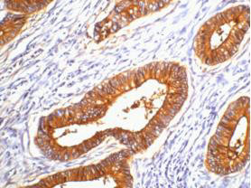
- Application Supplier NoteCH1A1A is a humanized, affinity matured, and stability-engineered version derived from the murine parental PR1A3 antibody, first disclosed in PMID: 2434440. This antibody is also basis of CEA TCB (RG7802), a CEA-CD3 T cell bispecific antibody currently in Phase 1/1b clinical trials (PMID: 27622073; 27117182; 26861458). This antibody was also used in the generation of cergutuzumab amunaleukin (CEA-IL2v, RG7813), a monomeric CEA-targeted immunocytokine, that comprises a single IL-2 variant (IL2v) moiety with abolished CD25 binding, fused to the C-terminus of a high affinity, bivalent carcinoembryonic antigen (CEA)-specific antibody devoid of Fc mediated effector functions. The binding affinity of this antibody for CEA was tested using ELISA. This antibody can also be used in the immunohistochemical analysis of CEA expression by various tumors (PMID: 28405498). This antibody was also used to study the surface expression heterogeneity of CEA in cells extracted from patient derived colorectal cancer organoids (PDOs) using flow cytometry (PMID: 30982469).
- ApplicationsFlow Cytometry, ELISA, ImmunoHistoChemistry
- Applications SupplierELISA; FC; IHC
- CertificationResearch Use Only
- ClonalityMonoclonal
- Clone IDCH1A1A (2F1, CH1A1A-2F1)
- Gene ID1048
- Target nameCEACAM5
- Target descriptionCEA cell adhesion molecule 5
- Target synonymsCD66e, CEA, cell adhesion molecule CEACAM5, carcinoembryonic antigen related cell adhesion molecule 5, meconium antigen 100
- HostHuman
- IsotypeIgG1
- Protein IDP06731
- Protein NameCell adhesion molecule CEACAM5
- ReactivityHuman
- Reactivity SupplierHuman
- Reactivity Supplier NoteThe original murine antibody PR.1A3 was generated by immunizing BALB/c mice with mucosal scrapings and cell membrane preparations from normal large intestine. Booster dose was given with same antigen or in one case boosting was also performed with HT29 colon carcinoma cells. CH1A1A was later one generated for affinity maturation and stability enhancement by phage display.
- Storage Instruction-20°C,2°C to 8°C
- UNSPSC41116161
Related products
Product group Antibodies
Anti-CEACAM5 AntibodyA100175
ApplicationsWestern Blot, ELISA
ReactivityHuman
- SizePrice
Product group Antibodies
Anti-CEACAM5 Antibody144-60045
ApplicationsWestern Blot
ReactivityHuman, Mouse
TargetCEACAM5
- SizePrice
Product group Antibodies

Anti-CEA [EB-011]Ab01002-1.1
ApplicationsELISA
ReactivityHuman
TargetCEACAM5
- SizePrice
Product group Antibodies
References
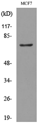
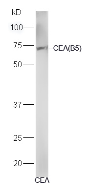
CEA(B5) Monoclonal AntibodyBSM-1624M
ApplicationsImmunoFluorescence, Western Blot, ELISA, ImmunoHistoChemistry, ImmunoHistoChemistry Paraffin
ReactivityHuman
TargetCEACAM5
- SizePrice

Product group Antibodies
Anti-CEA/CEACAM5 Antibody Picoband(r)A00356-2-CARRIER-FREE
ApplicationsFlow Cytometry, Western Blot, ELISA
ReactivityHuman
TargetCEACAM5
- SizePrice
Product group Antibodies
CEACAM5 Monoclonal AntibodyCSB-MA000218
ApplicationsELISA, ImmunoHistoChemistry
ReactivityHuman
TargetCEACAM5
- SizePrice
Product group Antibodies
ApplicationsWestern Blot
ReactivityMouse
TargetCEACAM5
- SizePrice
Product group Antibodies

ApplicationsFlow Cytometry, Western Blot, ImmunoCytoChemistry, ImmunoHistoChemistry, ImmunoHistoChemistry Frozen, ImmunoHistoChemistry Paraffin
ReactivityHuman
TargetCEACAM5
- SizePrice