


Immunohistochemical staining of human colon shows moderate cytoplasmic positivity in glandular cells.
Immunohistochemical staining of human colon shows moderate cytoplasmic positivity in glandular cells.
Anti-CEP63 Antibody
HPA058154
ApplicationsImmunoHistoChemistry
Product group Antibodies
ReactivityHuman
TargetCEP63
Overview
- SupplierAtlas Antibodies
- Product NameAnti-CEP63 Antibody
- Delivery Days Customer4
- ApplicationsImmunoHistoChemistry
- CertificationResearch Use Only
- ClonalityPolyclonal
- ConjugateUnconjugated
- Gene ID80254
- Target nameCEP63
- Target descriptioncentrosomal protein 63
- Target synonymsSCKL6, centrosomal protein of 63 kDa, centrosomal protein 63kDa, centrosome protein CEP63
- HostRabbit
- IsotypeIgG
- Protein IDQ96MT8
- Protein NameCentrosomal protein of 63 kDa
- Scientific DescriptionRecombinant Protein Epitope Signature Tag (PrEST) antigen sequence
- ReactivityHuman
- Storage Instruction-20°C,2°C to 8°C
- UNSPSC41116161
Datasheet
MSDS
Related products
Product group Antibodies
Cep63 Polyclonal AntibodyCAC09551
ApplicationsWestern Blot, ELISA, ImmunoHistoChemistry
ReactivityMouse
TargetCEP63
- SizePrice
Product group Antibodies
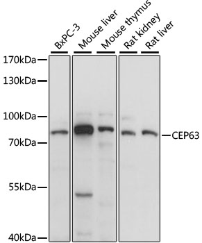
Anti-CEP63 AntibodyA91067
ApplicationsWestern Blot
ReactivityHuman, Mouse, Rat
- SizePrice
Product group Antibodies

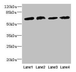
CEP63 AntibodyLS-C750486
ApplicationsWestern Blot
ReactivityHuman, Mouse, Rat
TargetCEP63
- SizePrice
Product group Antibodies
CEP63 AntibodyCSB-PA839376LA01HU
ApplicationsWestern Blot, ELISA, ImmunoHistoChemistry
ReactivityHuman, Mouse
TargetCEP63
- SizePrice

Product group Antibodies
Anti-CEP63 Antibody Picoband(r)A01661-1-CARRIER-FREE
ApplicationsWestern Blot, ELISA
ReactivityHuman
TargetCEP63
- SizePrice